Solitary Pulmonary Arteriovenous Malformation
(Click on small picutres to view larger images)
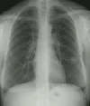
The patient's CXR demonstrates a nodular density in the left lower lung. A prominent vessel can be seen extending from the hilum towards the lesion.

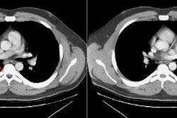
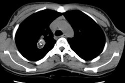
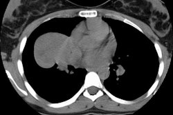
The patient's CT scan clearly demonstrates an enlarged pulmonary artery supplying the lesion and confirms it's vascular nature.

The pulmonary angiogram demonstrates a solitary feeding artery to the lesion.

The patient was treated by embolization. Post-procedure film demonstrates occlusion of the feeding pulmonary artery and no residual AVM.


