Esophageal Rupture:
Clinical:
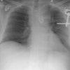
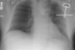
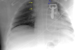
Esophageal rupture occurs in fewer than 1% of patients with blunt chest trauma. The sudden increase in intralumenal pressure associated with the trauma results in rupture, similar to Boerhave's syndrome). The rupture usually occurs in the left inferior posterior wall.X-ray:
Indirect signs of esophageal rupture include pneumomediastinum, left pneumothorax, and left pleural effusion. Pneumomediastinal air along the inferior descending aorta and left cardiovertebral angle of the diaphragm produce a characteristic "V" sign of Naclerio. The presence of a rupture can be confirmed with an esophagram using non-ionic contrast media.CT findings suggestive of esophageal perforation include wall thickening, a focal wall defect, and periesophageal fluid and air [1]. CT is highly sensitive for the detection of esophageal perforation and fluoroscopic esophagography is unnecessary following a negative CT exam [1].
REFERENCES:
(1) AJR 2013; Wu CH, et al. Esophagography after penumomediastinum without CT findings of esophageal perforation: is it necessary? 201: 977-984