Pulmonary Laceration:
Clinical:
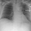
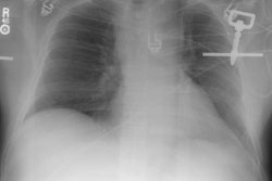
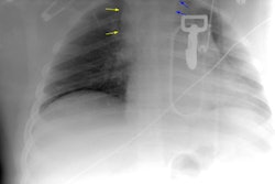
Pulmonary laceration represents a tear in the lung. It is typically the result of blunt high energy trauma or puncture wound and over half of the affected patients will have significant injury to another major organ system (pulmonary contusion results from a lesser magnitude of lung injury). Because of the normal elastic recoil of the tissue, the surrounding lung pulls back from the laceration and it will appear as a round or oval cavity (not a linear appearance) [4]. If the laceration fills with blood, a spherical hematoma forms. If it fills with air, a traumatic pneumatocele or air cyst forms. If both blood and air are present, an air-fluid level may be seen (hematopneumatocele) [3]. Over time the laceration becomes increasingly filled with blood [4]. Clearing is usually slow- with findings persisting for months or years. CT is superior to plain films in the detection of pulmonary lacerations [3]. Pulmonary laceration is common in children and young adults because they have greater flexibility of the chest wall, resulting in a higher likelihood of lung injury with blunt trauma [4].Four types of laceration have been described: Type I- compression-rupture injury- this is the most common type [4]. A direct compressive force results in lacerations in a deep portion of the lung [4]. Type 2- compression-sheer injury- a severe sudden blow to the lower hemithorax results in a sudden shift of the lower lobes across the spine [4]. These lacerations are seen in the paraspinal regions [4]. Type 3- rib penetration tear- are located in the periphery adjacent ot a fractured rib and an associated pneumothorax is generally seen [4]. Type 4 - adhesion tear- is a laceration in the region of a preexisting pleuropulmonary adhesion [4].
X-ray:
Immediately after trauma the radiographic evidence of pulmonary laceration is often obscured by the associated lung contusion. Acutely, the hematoma within the laceration produces a well circumscribed, homogeneous opacification (as previously mentioned, this finding may be partially obscured by overlying contusion). With clearing of the contusion the lesion will become more evident. Within several hours to a few days, round or oval air collections will begin to be identified within the lesion as the hematoma within the laceration is reabsorbed. This is called a post-traumatic pneumatocele or post-traumatic lung cyst. When the ruptured airspace is partially filled with blood an air-fluid level can be identified within the cyst (and the lesion may be confused with a lung abscess).REFERENCES:
(1) Semin Ultrasound, CT, MRI 1996; 17(2): 114-118
(2) Radiographics 1998; Van Hise ML, et al. CT
in blunt chest trauma: Indications and limitations. 18: 1071-1084
(3) Radiographics 1998; Kuhlman JE, et al. Radiographic
and CT findings of blunt chest trauma: Aortic injuries and looking beyond
them. 18: 1085-1106
(4) Radiographics 2008; Kaewlai R, et al. Multidetector CT of blunt thoracic trauma. 28: 1555-1570