Pulmonary Contusion:
Clinical:
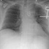
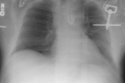
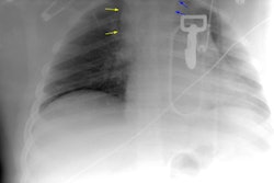
Pulmonary contusion is the most common lung injury from blunt chest trauma [5]. Pulmonary contusion is present in 30% to 70% of patients following blunt trauma and reflects the presence of edema and interstitial and intraalveolar hemorrhage. Hemoptysis will occur in up to 50% of cases, but dyspnea and hypoxia occur only with large contusions. An estimation of the severity of the contusion is significant clinically because it correlates with the degree of post-traumatic respiratory insufficiency. Mortality from pulmonary contusion is about 10%, but is as high as 70% in patients with extensive pulmonary injury. [1] All pulmonary contusions are present radiographically by 24 hours and generally resolve within 5-7 days [6]. Focal pulmonary opacities appearing 24 hours or more after injury suggest a diagnosis other than contusion (aspiration, pneumonia, or fat embolism) [5].X-ray:
CXR: Radiographically a contusion appears as an ill-defined, non-segmental pulmonary opacification. The contusion most commonly underlies the region of impact (90%), but contrecoup lesions may also be seen. A contusion is typically visible at presentation or within 6 hours (85%). The short time interval is helpful in differentiating contusions from fat emboli which may have a similar appearance, but are usually not apparent until one or more days following the injury. The infiltrate typically resolves over 2-5 days (complete clearing in 3-10 days [5,6]). Adjacent rib fractures are commonly seen.Computed tomography: CT is superior to plain films in detecting pulmonary contusions and a pulmonary contusion is highly unlikely in patients with a normal CT. On CT a contusion appears as an ill-defined area of consolidation or ground glass opacification adjacent to (or opposite- contrecoup) the area of trauma [2-4].
REFERENCES:
(1) AJR 1996; Song JK, et al. Diagnosis of pulmonary contusions and a bronchial laceration after a fall.167:1510 (No abstract available)
(2) Semin Ultrasound, CT, MRI 1996: Apr. 17 (2) : 114-118
(3) AJR 1997; Ground-glass opacity at CT: the ABCs. 169: 355-367 (No abstract available)
(4) Radiographics 1998; Van Hise ML, et al. CT in blunt chest trauma: Indications and limitations. 18: 1071-1084
(5) Radiographics 2008; Kaewlai R, et al. Multidetector CT of blunt thoracic trauma. 28: 1555-1570
(6) AJR 2009; Ho ML, Gutierrez FR. Chest radiography in thoracic polytrauma. 192: 599-612