Superior image quality gives flat panel digital x-ray the edge over screen-film and computed radiography (CR) systems when it comes to imaging the hand, according to a poster presentation at the 2001 RSNA meeting.
Researchers from Krakenhaus Dresden-Friedrichstadt in Dresden, Germany compared hand images obtained on 100 patients who presented for a rheumatologic baseline or follow-up exam. Half the patients had a randomly selected hand imaged with a Revolution XR/d general purpose flat-panel digital system (GE Medical Systems, Waukesha, WI). The unit was mounted on a radiographic table and had a 200-micron pixel size.
The other hand was imaged using extremity 100-speed screen film (Curix HT 1.0 G Plus, Agfa Healthcare, Ridgefield Park, NJ). The same DR system was used to image one hand of the remaining 50 patients while the other hand was scanned with the ADC Compact CR system (Agfa Healthcare).
All images were obtained using the same x-ray tube and generation, sans a grid with 100-cm source-to-detector distance, 50 kVp, and 2.5-3.2 mAs. The DR and CR images were printed on a laser printer. Using a 5-point scale, five radiologists scored image quality based on eight anatomical criteria:
- cortical structures of the unguiculares
- cortical structures of the metacarpal bones
- cortical-trabecular borders of the distal radius
- metacarpal-phalangeal joints
- periarticular soft tissues of metacarpal-phalangeal joints
- trabecular structures of two carpal bones
- periarticular soft tissues of the wrist
The radiologists also judged the images on overall quality.
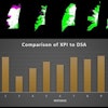
According to the results, all anatomical structures on the DR images were scored equal to, or better than, the screen-film and CR images. DR was graded superior by three of five readers for cortical structures and trabecular structures over screen-film. Four readers thought DR images were better for border structures, soft tissue, and overall image quality. The DR images fared as well in the same categories when compared to CR images, the authors reported.
"We found a significantly improved visualization of regions with thick or dense tissue, and of regions with thin or less dense tissue, which is enabled by the high dynamic range and linear characteristic curve of the digital system," Dr. Heike Pauls and co-authors wrote.
They also said they achieved good soft-tissue contrast through the DR system’s high Detective Quantum Efficiency and bit depth of 14 bits.
While the DR system has a relatively low resolution of 2.5 lp/mm, the authors still rated it as offering "consistently very good image quality for extremity exams."
By Shalmali Pal
AuntMinnie.com staff writer
November 26, 2001
For the rest of our coverage of the 2001 RSNA meeting, go to our RADCast@RSNA 2001.
Copyright © 2001 AuntMinnie.com