An artificial intelligence (AI) algorithm could boost the performance of x-ray for screening suspected thoracic aortic dissection in emergency department patients, a study published online December 19 in Scientific Reports indicates.
A group from Hanyang University in Seoul trained a convolutional neural network (CNN)-based algorithm to detect thoracic aortic dissection on chest x-rays. In a dataset of 204 patients with confirmed cases who presented to a local emergency department with chest pain, the model yielded an accuracy of 90%, they reported.
"Given the high severity and acuity of thoracic aortic dissection, early suspicion based on chest x-rays and CNN could accelerate the diagnosis and improve the prognosis," wrote corresponding author Tae Hyun Kim, PhD, of the university's Machine Learning Research Center for Medical Data.
Acute thoracic aortic dissection is a rare but life-threatening tear in the wall of the aorta. Blood surges through the tear, causing the inner and middle layers to separate or "dissect." The condition has a mortality rate of 50% within 48 hours if not diagnosed and treated properly.
Primarily, physicians use CT images to diagnose aortic tears, and this method is highly effective. But CT images may not be taken for hours after a patient presents in the emergency room, according to the authors. That's why chest x-ray is commonly used to rapidly differentiate causes of chest pain.
Still, the sensitivity of chest x-ray scans for acute thoracic aortic dissection is only 70%, which is not high enough given the fatality of aortic dissection, the authors wrote. In this study, they hypothesized they could improve this diagnostic accuracy through use of deep learning software.
The researchers culled 3,331 images (716 positive and 2,615 negative) from 3,331 patients treated at three hospitals in Seoul between 2003 and 2020. They chose a ResNet-18 neural network and trained it on a dataset of 1,972 images (507 positive cases) from one hospital, while a dataset of 1,155 images (155 positive cases) from a second hospital served as validation. For testing, they used a dataset of 204 images from the third hospital, 54 of which were positive for aortic dissection.
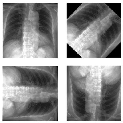
 Image preprocessing. Image courtesy of Scientific Reports through CC BY 4.0 International License.
The primary objective for the algorithm was to classify chest x-ray images into one of two categories: normal or aortic dissection. Training images were augmented via rotation, horizontal flipping, and vertical flipping, the authors noted; to evaluate the performance of the final deep learning model, they used the receiver operating characteristic curve (ROC) measure and calculated its area under the curve (AUC).
According to the findings, the algorithm achieved an AUC of 0.955 on the testing dataset. Ultimately, the analysis revealed the diagnostic accuracy of the deep-learning model was 90.2% for identifying images with thoracic aortic dissection.
But chest x-ray for this indication isn't quite ready to replace CT imaging, the authors wrote.
"Although the performance of the aortic dissection classification model was good, our observations are not sufficient to conclude that chest x-rays can be used to replace CT scans completely in patients with suspected aortic dissection," they noted.
The algorithm's performance represents an improvement over emergency radiologists' performance, according to Kim and colleagues: One study found a sensitivity of 86% and an accuracy of 60% among radiologists when diagnosing aortic dissection based on abnormal chest x-ray findings.
Additional studies are warranted, the group urged.
"In future works, the accuracy should be improved based on segmentation, and research directly applicable to clinical practice should be prioritized," the team concluded.