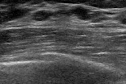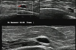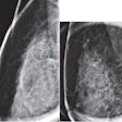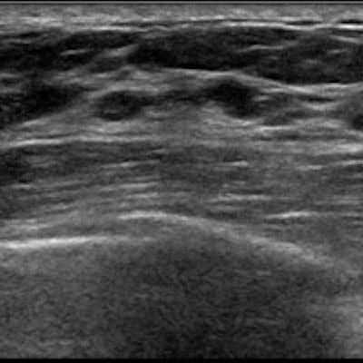
Ultrasound is more accurate for measuring the pathology size of breast tumors than mammography or MRI, according to a Canadian study published March 31 in the American Journal of Surgery.
Researchers led by Hannah Kapur from the University of British Columbia found that all three imaging methods overestimate tumors over 50 mm, while ultrasound and mammography underestimate tumors up to 20 mm.
"Concordance decreases with increasing size and lobular histology," Kapur and colleagues wrote. "Patients can be reassured that imaging size can be used dependably by surgeons to plan lumpectomy for clinical T1 tumors."
Total mastectomy or breast-conserving surgery are standard treatment methods for breast cancer. However, researchers wrote that overtreatment is a concern among surgeons and mastectomy rates are increasing. The European Society of Breast Cancer Specialists says that 85% of invasive cancers smaller than 30 mm should undergo breast-conserving surgery.
The researchers also wrote that as imaging methods improve, more and more of the detected breast cancers can't be found via clinical examination. Surgeons use quality indicators that involve the preoperative imaging size of tumors to select patients for breast-conserving surgery.
"It has been recognized that patients perceive mastectomy to be superior treatment, or they may want to avoid the possibility of subsequent procedures that may be necessary in the setting of breast-conserving surgery," the study authors wrote.
Kapur et al wanted to find out if preoperative tumor sizes from mammography, ultrasonography, and MRI could accurately inform surgical decision-making. They wrote that doing so could help with patient counseling and quality indicator recommendations.
The team identified 1,512 tumors among 1,502 patients who had surgery for invasive breast cancer between 2013 and 2017. Ultrasound was used on 1,251 patients, mammography on 922, and MRI on 75. Imaging reports came from 28 different imaging facilities.
The researchers compared preoperative imaging size with postoperative pathology sizes to look at correlation and agreement.
| Agreement between preoperative imaging size and postoperative pathology sizes from imaging methods | |
| Imaging modality | Agreement |
| Ultrasound | 0.628 |
| Mammography | 0.571 |
| MRI | 0.570 |
The study authors wrote that their findings suggest that ultrasound and mammography are effective at estimating postoperative pathology sizes for T1 and T2 tumors. Ultrasound underestimated T1 and T2 tumors while mammography underestimated T1 tumors and overestimated T3 tumors. Ultrasound also had the highest agreement with postoperative pathology sizes for T1 and T2 tumors, the team found.
"MRI has been found to overestimate tumor size and have the highest error rates and our results are consistent with this, although the number of patients having MRI in our study is small," the authors added.
However, the researchers wrote that the diversity of information for patients in the study makes it difficult to draw conclusions about what imaging to recommend preoperatively and when additional studies, such as MRI, would be useful.
"Although it would be ideal to have an algorithm that would identify the patients where imaging is not accurate, the results of this study have shown us that multiple factors need to be taken into consideration," they stated.


