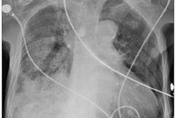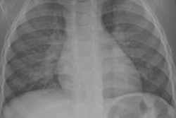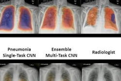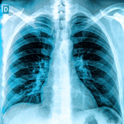
An AI model trained to detect pneumonia in chest x-rays of adults may also be valuable in pediatric patients ages 1 to 5, according to a recent study in the Journal of the American College of Radiology.
A team led by George Morcos, MD, of the University of Maryland in Baltimore, found that an AI model trained on adult x-ray images differentiated a significant number of normal versus pneumonia cases in a dataset of 5,856 pediatric chest x-rays.
"Adult-trained AI algorithms can provide a great starting point for AI applications in pediatric imaging to help address the significant disparity between adult and pediatric AI research," the group wrote.
The development of AI algorithms is booming in radiology, yet the authors noted that there is a scarcity of applications aimed for use with children. Of more than 200 commercial AI tools listed in a database managed by the American College of Radiology (ACR), only 4% are approved for children and none of them are specifically designed for thoracic imaging, Morcos and colleagues noted.
This could cause a significant health disparity in this vulnerable population, the authors suggested. Thus, in this study, they launched an initial effort to determine whether an algorithm trained to detect pneumonia in adults could also be used to detect pneumonia in pediatric patients.
The researchers used an AI algorithm called the "all" model from TorchXRayVision, an open-source software library for working with chest radiograph datasets and deep-learning models. Primarily, the models have been trained on adult datasets.
For data in this study, the researchers used a publicly available pediatric chest x-ray set developed at Guangzhou Medical Center in Guangzhou, China. The dataset contains 1,583 normal cases and 4,273 cases with pneumonia, differentiated as bacterial (n = 2,780) or viral (n = 1,493).
Overall, the algorithm identified 3,923 cases as pneumonia (67%) and 1,933 (33%) as normal. Further analysis showed the following:
- In the bacterial pneumonia group, the algorithm identified 2,734 cases as pneumonia (63%) and 1,629 (37%) as normal.
- In the viral pneumonia group, the algorithm identified 1,701 cases as pneumonia (55%) and 1,375 (45%) as normal.
"Although the performance of the algorithm was satisfactory for overall findings and in the bacterial pneumonia subset, there was decreased AI performance in the case of viral pneumonia," the authors wrote.
Ultimately, while a "satisfactory" performance may not measure up to the performance of the AI algorithm seen in adult patients, the results are encouraging in terms of leveraging adult-based algorithms and datasets for pediatric AI research, the authors noted.
"Given the relative scarcity of pediatric datasets and AI applications, we encourage attempting to leverage adult models and datasets to accelerate pediatric AI research and the implementation of its clinical applications," the group concluded.
The full article was published July 17 and is available here.