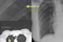
Computer-aided detection (CAD) developer Riverain Medical of Miamisburg, OH, has received clearance from the U.S. Food and Drug Administration (FDA) to market its SoftView enhanced chest imaging technology.
SoftView is designed to suppress ribs and clavicles on chest x-rays to produce a soft-tissue image that enhances clarity and the detection of lung nodules. The technology can be used on existing x-ray equipment to provide a soft-tissue image for digital chest x-rays.
SoftView delivers no additional radiation dose to the patient and does not require any additional patient procedures or specialized equipment.
Related Reading
Riverain scores Canadian sales deal, November 19, 2009
Riverain scores regulatory approvals, November 19, 2008
Road to RSNA, CAD, Riverain Medical, October 30, 2008
Riverain gets VA contract, July 23, 2008
Cleveland Clinic, Riverain to evaluate chest x-ray CAD, November 27, 2007
Copyright © 2010 AuntMinnie.com



![A normal mammogram confirmed by three-year radiologic follow-up illustrates reader-marked regions of interest (ROIs) during (A) unaided (round 1) and (B) artificial intelligence (AI)–assisted (round 2) reading. Each colored dot represents an ROI for recall by a human reader. Readers could mark more than one ROI per case, represented by multiple dots of the same color. During AI-assisted reading, the AI system displayed three visible prompts: two with suspicion of malignancy scores of 35% (left mediolateral oblique [L MLO] and craniocaudal [L CC]) and one with a suspicion of malignancy score of 10% (right craniocaudal [R CC]), shown as polygonal overlays. Without AI, six of 10 readers (60%) marked a false-positive ROI. With AI assistance, this fell to two of 10 (20%). R MLO = right mediolateral oblique.](https://img.auntminnie.com/mindful/smg/workspaces/default/uploads/2026/07/2026-07-14-radiology-mammogram-ai-auto-bias.H0bYO8QlWs.jpg?auto=format%2Ccompress&fit=crop&h=100&q=70&w=100)


![A normal mammogram confirmed by three-year radiologic follow-up illustrates reader-marked regions of interest (ROIs) during (A) unaided (round 1) and (B) artificial intelligence (AI)–assisted (round 2) reading. Each colored dot represents an ROI for recall by a human reader. Readers could mark more than one ROI per case, represented by multiple dots of the same color. During AI-assisted reading, the AI system displayed three visible prompts: two with suspicion of malignancy scores of 35% (left mediolateral oblique [L MLO] and craniocaudal [L CC]) and one with a suspicion of malignancy score of 10% (right craniocaudal [R CC]), shown as polygonal overlays. Without AI, six of 10 readers (60%) marked a false-positive ROI. With AI assistance, this fell to two of 10 (20%). R MLO = right mediolateral oblique.](https://img.auntminnie.com/mindful/smg/workspaces/default/uploads/2026/07/2026-07-14-radiology-mammogram-ai-auto-bias.H0bYO8QlWs.jpg?auto=format%2Ccompress&fit=crop&h=112&q=70&w=112)