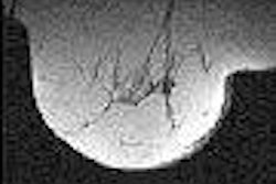
Computer-aided detection (CAD) firm Confirma has introduced the next generation of its CADstream breast MRI CAD system.
The new system features enhancements such as a streamlined portfolio for more comprehensive treatment planning and a multimodality-ready configuration that allows users to access, import, and display mammography and ultrasound studies. It also includes an adaptive registration (2D/3D) algorithm, according to Kirkland, WA-based Confirma.
In other Confirma news, the company said that over 200 imaging centers worldwide now use CADstream. In addition, Confirma said that it has received a Canadian medical device license, and has initiated commercial activities in Canada.
By AuntMinnie.com staff writers
May 26, 2005
Related Reading
Vital Images signs Confirma distribution deal, May 19, 2005
Confirma to unveil breast MRI coil at RSNA show, November 19, 2004
Confirma brings CADstream to Europe, October 29, 2004
Confirma hits CADstream mark, August 4, 2004
Copyright © 2005 AuntMinnie.com
![A normal mammogram confirmed by three-year radiologic follow-up illustrates reader-marked regions of interest (ROIs) during (A) unaided (round 1) and (B) artificial intelligence (AI)–assisted (round 2) reading. Each colored dot represents an ROI for recall by a human reader. Readers could mark more than one ROI per case, represented by multiple dots of the same color. During AI-assisted reading, the AI system displayed three visible prompts: two with suspicion of malignancy scores of 35% (left mediolateral oblique [L MLO] and craniocaudal [L CC]) and one with a suspicion of malignancy score of 10% (right craniocaudal [R CC]), shown as polygonal overlays. Without AI, six of 10 readers (60%) marked a false-positive ROI. With AI assistance, this fell to two of 10 (20%). R MLO = right mediolateral oblique.](https://img.auntminnie.com/mindful/smg/workspaces/default/uploads/2026/07/2026-07-14-radiology-mammogram-ai-auto-bias.H0bYO8QlWs.jpg?auto=format%2Ccompress&fit=crop&h=100&q=70&w=100)


![A normal mammogram confirmed by three-year radiologic follow-up illustrates reader-marked regions of interest (ROIs) during (A) unaided (round 1) and (B) artificial intelligence (AI)–assisted (round 2) reading. Each colored dot represents an ROI for recall by a human reader. Readers could mark more than one ROI per case, represented by multiple dots of the same color. During AI-assisted reading, the AI system displayed three visible prompts: two with suspicion of malignancy scores of 35% (left mediolateral oblique [L MLO] and craniocaudal [L CC]) and one with a suspicion of malignancy score of 10% (right craniocaudal [R CC]), shown as polygonal overlays. Without AI, six of 10 readers (60%) marked a false-positive ROI. With AI assistance, this fell to two of 10 (20%). R MLO = right mediolateral oblique.](https://img.auntminnie.com/mindful/smg/workspaces/default/uploads/2026/07/2026-07-14-radiology-mammogram-ai-auto-bias.H0bYO8QlWs.jpg?auto=format%2Ccompress&fit=crop&h=112&q=70&w=112)