A tracer kinetic model can enable quantitative measurement of cardiac amyloidosis burden and assessment of treatment response, according to a study presented at the annual Society of Nuclear Medicine and Molecular Imaging (SNMMI) meeting in Chicago.
After assessing a number of methods for quantifying F-18-flutemetamol myocardial uptake on PET exams in patients with transthyretin (ATTR) cardiac amyloidosis, researchers led by Qiong Liu, a doctoral student at Yale University in New Haven, CT, found that a two-tissue reversible compartment model produced the best results in patients with this rare, life-threatening disorder.
"These findings suggest that commonly employed semiquantitative methodologies, such as standardized uptake values used for [F-18 FDG], are not optimal for [F-18-flutemetamol]," Liu said in a statement from the SNMMI. "Using parametric modeling to fully quantify volume of distribution may provide clinicians with a more accurate and robust method for assessing the presence and progression of amyloid plaques in the myocardium, ultimately improving patient care."
Although F-18 flutemetamol PET imaging has shown potential for detecting ATTR cardiac amyloidosis, static PET data analysis methods for these exams are time-sensitive due to patient-dependent tracer washout from the myocardium.
"As a result, these methods can only provide physicians with a limited read-out," Liu said. "New methods to obtain fully quantitative myocardial volume of distribution with this tracer are needed."
In an effort to determine the optimal method for quantifying F-18 flutemetamol myocardial uptake, the researchers performed kinetic modeling on dynamic PET scans of six ATTR cardiac amyloidosis patients. After dynamic images were reconstructed, the team then tested several compartmental models and analyzed parametric images of the volume of distribution.
A two-tissue reversible compartment model was the winner. The simplified graphical Logan plot identified the volume of distribution parameter generated from the first 30-minute dynamic scan was a quantitative parameter deemed by the researchers to be potentially useful for diagnosing and evaluating treatment in patients with ATTR cardiac amyloidosis.
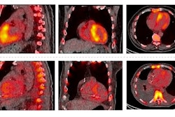
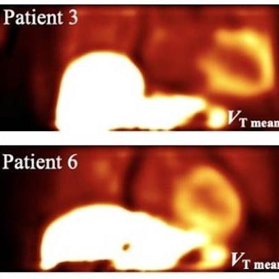
 VT images for six patients generated from 30 minutes of dynamic data. Mean VT values of myocardium are shown on the bottom right. Images courtesy of Qiong Liu and the SNMMI.
To use any new tracer optimally, it's especially important to understand the fundamental kinetics of tracers in the target organ, the researchers emphasized. They also noted that similar approaches using dynamic and parametric imaging may be beneficial for other tracers as well.
The study underscores the importance of understanding the fundamental kinetics of tracers in the target organ of interest, which is especially important to the optimal use of any new tracer, according to the researchers. Similar approaches using dynamic and parametric imaging may also be beneficial for other tracers, they said.