Dear AuntMinnie Member,
Our most popular story this week on AuntMinnie.com featured recent research revealing that interval breast cancers have less-favorable molecular features compared with those detected on screening mammography.
Interval breast cancers were found to often have higher tumor and nodal staging, frequently be triple-negative, and have high Ki67 proliferation indices. What's more, they're more regularly associated with dense breast tissue, according to the researchers.
You can view our coverage by stopping by our Women's Imaging Community. While you're there, you can also check out other new stories, including how a nomogram based on radiomics and other ultrasound features was recently deemed helpful for assessing axillary lymph node status in patients with early invasive breast cancer.
Real-world AI performance
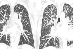
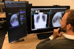
How well does artificial intelligence (AI)-based computer-aided detection (CAD) software perform in the real world on chest x-rays? Pretty well, if the experience of Seoul National University Hospital in Korea is any indication. After an AI-based CAD algorithm was embedded into their hospital's screening workflow, radiologists doubled the number of nodules that needed follow-up.
In other stories featured this week in your Digital X-Ray Community, researchers have trained an AI algorithm to estimate sex on digital x-rays using a skeleton collection belonging to the Smithsonian's National Museum of Natural History. In their proof-of-concept study, they found that the algorithm could accurately estimate sex 90% of the time, showing the potential for AI to aid in forensic analysis of skeletal remains.
And speaking of AI, internal medicine and emergency medicine physicians benefited more in a recent study than radiologists after receiving explainable AI advice when reading chest x-rays. But radiologists did have higher diagnostic confidence after accessing the AI advice.
Thyroid nodule risk stratification
A number of risk-stratification systems are available for classifying thyroid nodules, but there is one clear winner, according to a new study. The American College of Radiology Thyroid Imaging and Reporting and Data System outperformed five other systems in a meta-analysis that encompassed nearly 50,000 patients.
Also, the combination of historical clinical data and quantitative ultrasound analysis was found to better identify pregnant women at risk for spontaneous preterm birth, according to another highly viewed article in our Ultrasound Digital Community.