HONOLULU - Three-dimensional transcranial power Doppler imaging (3D-PDI) has shown promise as a rapid, noninvasive screening method for visualizing ruptured intracranial aneurysms in the emergency room, researchers reported Tuesday at the American Academy of Neurology (AAN) meeting.
In some cases, the technique may yield more useful clinical information than conventional x-ray angiography, according to Dr. Fabienne Perren and colleagues at the Neurology Clinic of Mannheim in Germany. The clinic is part of the University of Heidelberg.
In a comparison of 3-D-PDI with angiography to visualize and characterize acutely bleeding intracranial aneurysms, the two methods produced similar results. However, in several cases, the ultrasound images obtained provided better visualization of thrombosed aneurysms.
"In the last several years, there has been increasing interest in noninvasive assessment of intracranial aneurysm with ultrasound," Perren said. "Preoperative transcranial color-coded Doppler flow imaging allows characterization and localization of aneurysms as well as identification of vessels potentially threatened by clipping, whereas
preoperative Doppler sonography has been shown to be an effective additional alternative to preoperative angiography for the assessment of vessel patency in aneurysm surgery."
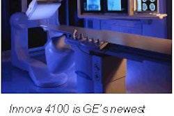
In the study, seven patients with acute subarachnoid hemorrhage, diagnosed with CT, underwent 3-D-PDI and x-ray angiography. The ultrasound exams were conducted on a phased-array system (ATL HDI 5000, Philips Medical Systems, Andover, MA).
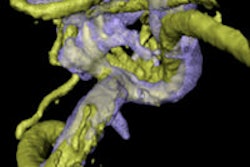
Unenhanced 3-D ultrasound images were acquired through each temporal bone window, using a slow sweep through the circle of Willis beginning at the vascular structures nearest the base of the skull and ending with the probe angled toward the parietal lobe. In all cases the volume scan was acquired within eight minutes. A dynamic real-time display of the reconstructed volume data was used to locate and characterize bleeding aneurysms.
3-D-PDI diagnosed four aneurysms of the anterior communicating artery, two aneurysms of the right middle cerebral artery, and one aneurysm of the left posterior communicating artery. In two aneurysms, thrombosis was clearly depicted by 3-D-PDI. The diagnosis of intracranial aneurysm was angiographically confirmed in all cases with identical location and size.
The main advantage of 3-D-PDI is its ability to pinpoint the basal arteries and circle of Willis, Perren said. "Moreover, it allows demonstration of intracranial vascular anatomical relationships by spatial representation and details that cannot be appreciated on 2-D images alone," she added. Other benefits of the technique include less operator dependence, as well as rapid volume reconstruction and rendering techniques.
In an interview with AuntMinnie.com, Perren said further studies are needed to improve the sensitivity of 3-D-PDI for the detection of intracranial aneurysms, and to determine if 3-D-PDI can sufficient characterize bleeding aneurysms. Such information would allow faster neurosurgical intervention. She also cautioned that the technique cannot be performed in the absence of a sufficient temporal bone window.
By Jill SteinAuntMinnie.com contributing writer
April 2, 2003
Related Reading
Pseudoaneurysm size affects success of thrombin injection with US, April 1, 2003
Transthoracic ultrasound detects adequate reperfusion after anterior MI, October 30, 2002
Quick ultrasound screening for AAA recommended in older men, September 27, 2002
Cuban radiologists rely on transcranial ultrasound, July 4, 2002
Copyright © 2003 AuntMinnie.com