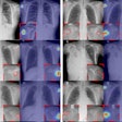
Dialing down the entrance skin dose while maintaining image quality is a main objective of x-ray research. A group from Belgium, working with a combined cesium iodide fine-needle scintillator and an amorphous silicon flat-panel DR system, believes it has achieved that goal.
According to results presented at the 2002 RSNA conference by a research team from Ghent University’s department of radiology, a DR system was able to achieve skin doses one-third to one-half the dose rate of conventional film or CR systems, respectively, in chest posteroanterior (PA) and lateral radiographic views.
"Our aim was to compare the patient entrance skin dose in three chest imaging systems: a full-digital amorphous silicon flat-panel detector, a conventional dedicated film-screen system, and a storage phosphor computed radiography system," said Dr. Peter Smeets, who delivered the group’s findings.
The researchers used DR technology from Trixell of Moirans, France, a joint-venture firm owned by Thales Electron Devices (51%) of Velizy, France; Philips Medical Systems (24.5%) of Andover, MA; and Siemens Medical Solutions (24.5%) of Erlangen, Germany. For the conventional film-screen system, the team used Siemens Thoramat equipment. The CR portion of the study was conducted on a DirectView CR 900 system from Eastman Kodak Health Imaging of Rochester, NY.
The researchers compared images taken from three groups of 100 patients who had been matched for age, gender, and body mass index. Each patient group was examined on the DR, conventional film, or CR system.
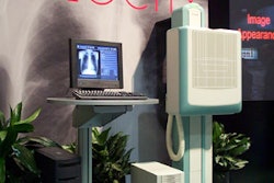
The DR system featured a 43 x 43-cm (17 x 17-inch) image receptor with a 3K x 3K detector surface, a 14-bit dynamic range, and a 143-micron pixel pitch. The cesium iodide fine-needle scintillator sat on top of the flat-panel detector and gave the unit a total weight of approximately 20 kg (44 lbs.) Smeet said.
"We adjusted the automatic exposure control on the flat-panel system to a 400-speed-class system to match the film-screen and CR systems’ performance," he said.
The team set the exposure specification for PA and lateral views to 125 kVp and used a 180-cm (71-inch) screen-focus distance to conduct its DR chest studies. The DR and CR images were transferred to a PACS workstation (MagicView, Siemens, and MasterPage, Kodak, respectively) for display on a 1.3K x 1.6K monitor (Siemens and BarcoView of Kortrijk, Belgium). The film-screen images were read off a lightbox.
Image quality for all three systems was evaluated by five radiologists using the European Quality Criteria for Chest Radiography (European Guidelines on Quality Criteria for Diagnostic Radiographic Images, Rep. EUR 16260, 1996, Office for Official Publications of the European Communities, L-2985 Luxembourg).
"Our conclusion was that the DR images were at least equal and actually better than the conventional film and CR performance," Smeets said.
Each patient had 24 calibrated thermoluminescent dosimeters (Harshaw TLD-100; Thermo Electron, Waltham, MA) attached at six defined and reproducible locations on the back and right side, Smeet said. After each view had been performed, the dosimeters were analyzed by a Harshaw 3500 manual TLD reader.
According to the results, the dose rate was lower in both views for the DR system than for the film-screen or CR system. The entrance skin dose rate for the PA view measured 66.8 μGy for the DR system, 199 μGy for the film-screen system, and 155 μGy for the CR-system. For the lateral view, the doses were 346.7 μGy, 1286.2 μGy, and 733.6 μGy for the DR, film-screen, and CR systems respectively.
"Overall, the DR system permits a significant reduction in radiation exposure compared with conventional film-screen or CR of approximately one-third to one-half, respectively, with no loss of image quality," reported Smeets.
A recent study conducted by Dr. Hakan Geijer at the department of radiology at Orebro Medical Centre Hospital in Orebro, Sweden, confirmed the efficiency of lower dose rates in DR versus film-screen or CR.
"The flat-panel detector had equal image quality at less than half the radiation dose compared with storage-phosphor plates. The difference was even larger when compared with film, with the flat-panel detector having equal image quality at approximately one-fifth the dose. The flat-panel detector has a very favorable combination of image quality versus radiation dose compared with storage-phosphor plates and screen-film," he wrote (European Radiology, September 2001, Vol. 11:9, pp. 1704-1709).
The lower dose rate potential for DR systems versus film-screen cannot be applied across the board to all radiographic studies. Writing in a supplement to Acta Radiologica (March 2002, Vol. 43:427, pp. 1-43) Geijer noted that the radiation dose for digital exposure was nearly twice as high as the film-screen method at a comparable image quality in scoliosis radiography.
"However, if the digital exposure protocol was optimized to a considerably lower dose with a slightly lower image quality, the flat-panel detector yielded a superior image quality at a lower dose than both storage-phosphor plates and screen-film," he wrote.
By Jonathan S. BatchelorAuntMinnie.com staff writer
January 30, 2003
Related Reading
Worrisome radiation dose seen in CT lung screening, follow-up, December 23, 2002
No compromise in breast image quality with lower FFDM dose, December 13, 2002
Even moderate doses of radiation may cause nervous system tumors, October 16, 2002
Technique and tools can reduce skin dose during interventional procedures, October 11, 2002
Study cuts radiographs and dose from colon exam, September 13, 2002
Copyright © 2003 AuntMinnie.com

