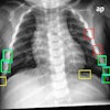
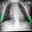
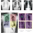
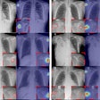
Although digital storage phosphor x-ray systems are capable of producing either 1,760 x 2,140 (2K) or 3,520 x 4,280 (4K) matrices for image acquisition, using the higher 4K resolution may not provide a clinically relevant increase in image quality. Radiologists from the department of diagnostic radiology at the University of Vienna, Austria, compared diagnostic reads on chest radiographs performed at both matrix sizes, and found no significant differences.
"The question is, is it necessary to really use the 4K matrices for the examination of chest radiographs? These large data sets put an increased workload on PACS and workstations, and we weren’t sure that the increased spatial resolution translated into additional diagnostic information," said Dr. Mathias Prokop, who presented the research team’s work at the 2001 RSNA meeting in Chicago.
The group obtained 100 clinical posteroanterior chest radiographs from 70 patients, covering a broad range of pathologies. The images were taken at 2K and 4K resolution, using dual-scanning storage phosphor plates. With the exception of the matrix size, all the images used the same full-size format, processing algorithm, and laser printing, Prokop said.
The images were blinded as to resolution, and then independently compared, side by side, by three radiologists. They used a three-point scale of better, equivalent, or worse to grade the comparisons. The team conducted two reading sessions. In the first, the radiologists viewed the whole image; in the second session, they chose various regions of interest for diagnostic reading.
Image assessment was based on four criteria. The radiologists first graded the visibility of the anatomic details of the vascular and bronchial structures, pleural interface, mediastinal lines, and ribs. They subsequently noted the pathology represented on each image, and assigned what they believed to be the correct matrix size. Finally, the researchers were asked to define the diagnostic impact of potential image differences.
None of the radiologists found additional diagnostic information using the 4K matrix size, Prokop reported. The researchers also found that the readers were unable to assign the image matrix in 56% of the images, and assigned it correctly in only 17% of the radiographs. Whether the whole image or only a region of interest was assessed at either matrix size (10% for the whole image and 4% for the region of interest), the results did not differ significantly.
"There was no appreciable difference for the assessment of mediastinal structures in 62% of the images, retrocardiac structures in 44%, pleural interfaces and ribs in 74%, and intrapulmonary structures in 58%," Prokop said.
He also noted that in the same four anatomic areas, there was no significant difference between the numbers of images that were assigned correctly or incorrectly to either image matrix.
"As a result of our studies, we deduced that a high-resolution 4K image does not provide a diagnostically relevant increase in image quality as compared to a 2K matrix for storage phosphor chest radiography," Prokop said.
By Jonathan S. BatchelorAuntMinnie.com staff writer
January 15, 2002
Related Reading
Flat-panel combo provides less noise, lower dose, November 29, 2001
Digital x-ray continues to make inroads, November 25, 2001
CR, DR vendors debate digital x-ray methods at SCAR meeting, June 4, 2000
Copyright © 2002 AuntMinnie.com


