Mammography remains the primary screening modality for breast cancer. For women with dense breasts, however, ultrasound is increasingly used as an adjunct diagnostic tool. A new company called U-Systems hopes to apply computer-aided detection (CAD) technology to breast ultrasound.
The San Jose, CA, company was founded by radiology veteran Bob Wang, who also founded CAD pioneer R2 Technology. The two companies said in a December 2000 press release that they would be working together "to explore the integration of CAD technology into ultrasound for breast cancer detection." The firms continue to remain close and collaborate on technology development. Although Wang resigned as chairman of R2 in 2001, he remains one of the firm's largest shareholders and serves as an advisor.
A former R2 executive -- Skip Mendelson-- serves as senior vice president. The majority of USI's technical staff were recruited from companies such as Diasonics and GE, Wang said.
USI’s core products are CAD tools for ultrasound breast cancer detection and image-guided breast biopsy. Wang also acquired ultrasound vendor Acoustic Imaging, taking over AI’s basic platform and building a new box around it. Changes also were made to the AI software and hardware architecture. Acoustic Imaging was originally a general ultrasound manufacturer, but had switched to breast imaging. Some 1,300 AI legacy systems are still in use.
Although USI’s newly dubbed Vista platform could function as a general ultrasound system, the multimodality companies have pretty much cornered that market, so USI is carving out a niche, according to the company.
USI is developing its architecture and software applications for dedicated breast imaging, because ultrasound can effectively distinguish between solid masses and cysts. The Vista CAD tools include Contour, CXR, Aspect, and Needle Site.
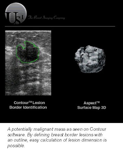
- Contour is software that defines breast lesion borders with a contouring outline. Malignant masses tend to have irregular borders, while cysts have a smoother outline. The outline also allows easy calculation of lesion dimensions.
- CXR is designed to mimic a chest x-ray image. Multiple planes of images are compressed to two dimensions, resembling a mammogram, and the radiologist can compare the two types of images.
- Aspect is a 3-D surface-map rendering that shows lesion surface characteristics. It can be used for needle visualization during biopsy procedures.
- Needle Site locates the biopsy needle in relation to the mass, and puts down track lights to show where the needle is and where it will go.
USI has a 510(k) on the Vista platform, while clearances are pending on Contour, Aspect, and Needle Site. The company expects to file this month on CXR. The vendor plans to show the entire system at the American College of Surgeons meeting in Boston, and at the American College of Radiology's April meeting in Dallas.
When asked if USI was working on integration of Aspect and Needle Site, so that a physician doing a breast biopsy could actually visualize the needle approaching and entering the 3-D rendering of a mass, Wang laughed, but wasn't about to give away his plans.
"That has been requested by physicians as something that would be useful to them, and we only take their suggestions into consideration," he said. "Whatever we’re working on, we won’t be talking about that at this point."
Making radiologists more productive
Bob Wang has made mammography the main focus of his career. Born in Beijing, he received his B.A. from Rensselaer Polytechnic Institute in Troy, NY, and his master’s in physics from the Massachusetts Institute of Technology (MIT) in Cambridge.
His first venture was International Medical Technology, a company founded in the 1970s to develop rare-earth screens for x-ray film. Up to that point, mammography had been done with chest photographic film whose sensitivity to x-ray was poor. With the introduction of rare-earth film screens, which intensified x-rays, dosage could be cut significantly and mass low-dose screening procedures became possible.
Wang sold that company to 3M, while he also licensed the procedure to Kodak. The rare-earth system became standard, and Wang made his fortune. His involvement with R2 Technology began in the early 1990s, when he became fascinated with the CAD technology being developed at the University of Chicago.
 |
The major modality vendors were skeptical of CAD, Wang said, so he obtained a license from the university in exchange for equity in the new company. It took about a year to assemble a team, raise venture money, and negotiate the final license agreement. R2 was incorporated in 1993, and its ImageChecker CAD system received premarket approval (PMA) in 1998.
Wang started USI in 1997 and resigned his R2 board position last year in order to devote more time to USI and another venture -- a company called Gazillion Bits -- developing photonics for applications to data communications. He is president and chairman of both companies.
Wang’s vision is the application of CAD to virtually all imaging modalities. He sees the technology primarily as a productivity tool, one that could play a role in alleviating the shortage of radiologists.
"If you look at what physicians today do, more than 90% of the images they look at do not contain any abnormalities," Wang said. "Not knowing where the abnormalities are, they have to search carefully at every image. I think that’s a total waste of time. So CAD eventually would have to be used to reduce, to help remove those normal images so they don’t have to spend so much time. Eventually, CAD will be shouldering that kind of work. This doesn’t just apply to mammography; it applies to all imaging -- CT, ultrasound, everything."
When CAD was first seen as a diagnostic tool, some radiologists feared computers were threatening to take away the human component. Thus, CAD went from being "computer-aided diagnosis" to "computer-aided detection," or an adjunct to the radiologist’s evaluation of an image.
"At last year’s RSNA, CAD was the main theme, so now CAD is mainstream. R2 was the hottest booth on the floor, and virtually every company said they are either developing or have some form of CAD. It’s a technology that 10 years ago nobody wanted, and all of a sudden now everybody’s embracing it."
For now, CAD is used only to reduce the incidence of gross errors and oversights that are the result of looking at so many normals, Wang said. By the time abnormalities appear, they may get missed.
"If every other case was a cancer, nobody would miss cancer," he said. "If a cancer is salted away in every 300 cases, then it’s very easy to overlook."
Wang believes CAD will become indispensable in radiology. USI’s plan is to introduce the same x-ray productivity tools developed at R2 to ultrasound.
"It won’t replace the radiologist. It will make the radiologist more productive," he said.
By Robert BruceAuntMinnie.com contributing writer
February 27, 2002
Related Reading
R2 CAD unit approved for use with GE's FFDM system, December 6, 2001
R2 Technology installs 200th mammography CAD system, October 22, 2001
New ultrasonic approach focuses on breast imaging , November 27, 2001
Copyright © 2002 AuntMinnie.com