VIENNA - Accurate staging of rectal carcinoma is essential for optimizing patient management, and ultimately, providing the best hope of long-term survival. Radiologists currently use tools such as MRI, CT, and endoluminal ultrasound to decide whether radiation and chemotherapy would be helpful, for example, or whether laparaoscopic rather than open resection is possible.
In Friday’s scientific sessions of the European Congress of Radiology, Dr. Jens Stollfus from the Technical University of Munich in Germany concluded that MR is superior to both ultrasound and CT for determining local tumor invasion in patients with advanced rectal carcinoma.
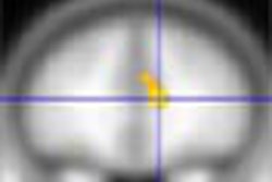
The group examined 103 patients (76 male, 27 female, mean age 64 ± 3 years), all of whom had initial presentation with tumors greater than or equal to T3 at initial presentation. Following bowel preparation and gadolinium enhancement, phased-array T1- and T2-weighted MRI was performed in all patients using a Gyroscan 0.5-tesla scanner (Phillips Medical Systems, Best, the Netherlands, equipped with a body coil, Stollfus said.
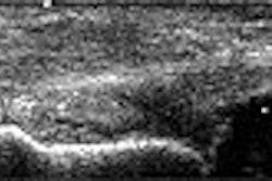
Then, multislice CT was performed in all patients 90 seconds following the administration of butyl-scopolamide IV contrast, using a protocol that included 120-140 mAs, pitch 5, and 8-mm slice thickness. Finally, all patients underwent endoluminal ultrasound performed by experienced surgeons.
The images were analyzed by consensus reading of two experienced radiologists, and classified according to TNM categories, relying primarily on local tumor invasion and the presence of lymph-node metastases as the main parameters. Finally T1-weighted and T2-weighted MR exams were compared in a subgroup of 43 patients. Following resection, histopathology was available for all results.
"The presence of intraluminal mass with mesorectal involvement was a major determinant in subsequent data analysis," Stollfus said.
According to the results, MRI was more accurate than either CT or ultrasound for the prediction of local tumor invasion (p < 0.01), Stollfus said.
MRI had a sensitivity of 84% for the detection of tumor invasion, compared with 69% and 71% for CT and EUS, respectively. In specificity, MRI achieved 68%, compared with 55% for CT and 28% for ultrasound.
MRI’s positive predictive value was 77%, compared with 65% for CT and 61% for EUS. Negative predictive value was 77% for MRI and 41% for EUS.
However, CT tended to be more specific regarding the prediction of nodal status (p < 0.05). And the group found no significant difference between contrast-enhanced and unenhanced MRI for the detection of local tumor invasion.
"MRI is superior to both CT and ultrasound for the prediction of local tumor invasion in patients with advanced rectal carcinoma, and may replace CT as the primary staging modality," he concluded, noting that lymph node characterization may improve with the development of USPIO-based MR contrast agents.
Following the talk, one audience member suggested that a 90-second delay following contrast administration may have been too long to achieve optimal sensitivity in CT, because such delays preclude the evaluation of tumors inside the rectal wall during the arterial phase. Another observer suggested that the use of multiplanar reconstruction may also improve CT results.
By Eric BarnesAuntMinnie.com staff writer
March 7, 2003
Related Reading
New colorectal cancer screening guidelines issued, February 4, 2003
In a screening population, virtual colonoscopy finds significant polyps, January 7, 2003
Medicare coverage has not increased colorectal cancer screening rates, December 30, 2002
Copyright © 2003 AuntMinnie.com