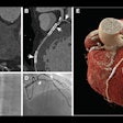
MIAMI BEACH, FL - Investigators from Michigan have developed what they consider to be an excellent protocol for a cardiovascular PET/CT exam. They discussed their technique Tuesday at the American Roentgen Ray Society (ARRS) meeting.
The goal is achieve accurate, rapid (one-hour exam) high-quality images from the combination of PET myocardial perfusion exams and CT angiography, said Dr. Kevin Berger.
Berger and his co-authors are from the departments of radiology and cardiology at Michigan State University in East Lansing, as well as GE Healthcare in Waukesha, WI. GE's Discovery ST scanner and its HeartFusion software for data display were used for this research.
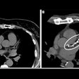
For the study, 16 patients underwent stress and myocardial perfusion stress scans with 20 mCi doses of 13N ammonia. Serial dynamic and gated myocardial perfusion PET images were obtained. Immediately following the PET scans, CT angiography (CTA) was performed using 100 cc of nonionic contrast (300 mg/ml) and 50 cc of saline, injected with a dual barrel at 4 cc/sec. An eight-slice CT scanner was used (Michigan State will be upgrading to a 16-slice scanner in June, Berger said).
The authors found that appropriate time-activity curves could be generated from a four-minute dynamic acquisition sequence (24 10-second frames). By collecting gated images (10 minutes or longer) immediately following dynamic image acquisition, they could diagnostically assess wall motion, Berger reported. Summed images of the bins allowed diagnostic interpretation of regional myocardial perfusion in all 16 cases.
The group then went a step further to reduce scan time, developing an image subtraction method for correcting background activity. They found that varying the scan intervals (25-50 minutes) on measured background activity produced decent diagnostic images, and that background subtraction was mandatory for PET stress/rest scan intervals of less than 40 minutes.
While their results were successful, Berger warned that "the equipment needs (for myocardial PET/CT) are extensive. You need a PET/CT scanner. There's extensive hardware and software for processing." In addition to extensive equipment needs, "personnel training is required to perform these combined exams."
An audience member asked if the group had encountered any co-registration artifacts. Berger said patient motion between rest and stress did result in falsely decreased perfusion in the anterial wall. The best way to prevent such a glitch would be to repeat the attenuation exams to check for changes brought on by motion, he said.
Session moderator Dr. Philip Costello praised the research as "the future of cardiac imaging." He also asked Berger about the degree of collaboration with the cardiologists.
"They've been fantastic," Berger replied. "They monitor the ECG exams and the radiologists read all the PET/CT studies. We discuss all of our exams together. (The cardiologists) wish we could do more of these types of exams."
By Shalmali Pal
AuntMinnie.com staff writer
May 5, 2004
Related Reading
Slower CT may be better in cardiac PET/CT, March 31, 2004
Gated PET offers one-stop assessment of multiple myocardial segments, June 23, 2003
Along with SPECT, PET and MRI find niche in myocardial assessment, October 21, 2002
Copyright © 2004 AuntMinnie.com


