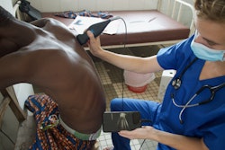
Training can help novice ultrasound users in the emergency department to successfully guide nerve blocks in patients with hip fractures, suggest findings published December 15 in JAMA Network Open.
This knowledge-to-practice training intervention improved nerve block uptake in hip fracture patients, with nerve blocks being quick, safe, and mostly effective. It also reduced delirium among patients, wrote a team led by Jacques Lee, MD, from the Schwartz/Reisman Emergency Medicine Institute at Sinai Health in Toronto, Ontario, Canada, and colleagues.
“The additional evidence provided by this study that regional anesthesia reduces incident delirium should challenge emergency department physicians and decision-makers to question whether [point-of-care ultrasound]-guided regional anesthesia should become the standard of care in their jurisdiction,” the Lee team wrote.
Delirium complicates between 20% and 62% of annual hip fractures. Previous studies suggest that point-of-care ultrasound (POCUS) could reduce delirium. However, the use of regional anesthesia for hip fractures varies by practice. Prior research also suggests that inadequate training and perceived time to perform blocks are the most common barriers.
Lee and co-authors developed a knowledge-to-practice intervention to train and encourage emergency department physicians to perform POCUS-guided regional anesthesia. For their study, the researchers assessed whether this approach could increase POCUS use by emergency department physicians and whether this affected the incidence and duration of delirium in older people with hip fractures.
The randomized clinical trial took place at seven academic emergency departments from four provinces in Canada. It included physicians working at least one shift per week who did not regularly use ultrasound for guiding anesthesia and patients ages 65 and older with a hip fracture who were not delirious on arrival.
The two-hour structured training session focused on fascia iliaca blocks. This was followed by a competency assessment, with a procedure bundle and email reminders provided to the participants.
Final analysis included 208 emergency department physicians who assessed 694 eligible patients with hip fractures (median age, 81) completed training. Among these patients, the study analyzed 248 patients prior to training per intention-to-treat analysis and 446 patients after training (intervention group).
The team reported the following findings:
The training led to increased nerve blocks, from six of 264 patients (2.2%) to 236 of 446 patients (52.9%) after training.
After adjustment, delirium risk decreased in the intervention group (odds ratio, 0.72). However, the average number of days of delirium was not reduced.
Among 186 nerve blocks with pain effectiveness data, 107 were effective with greater than 50% pain reduction.
The training led to a median nerve block procedure time of 15 minutes and one adverse event, a minor hematoma, was recorded.
The study authors highlighted that their results confirm the safety and effectiveness of their training program.
They called for future trials to confirm the impact of POCUS-guided regional anesthesia on delirium and explore the impact of improved early nerve block effectiveness on delirium risk.
“This may include training innovations to improve nerve block quality, duration, and uptake and exploring new approaches, such as pericapsular nerve group block and supra-inguinal approaches,” the authors wrote. “Finally, collaborations among the emergency department and anesthesia, orthopedic, and perioperative medicine departments are needed to improve nerve block rates.”
Read the full study here.