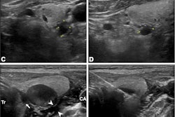
Outcomes over 10 years suggest that image-guided thermal ablation is a safe and effective option for patients with papillary thyroid carcinoma, according to a study published November 7 in JAMA Otolaryngology-Head & Neck Surgery.
The emerging procedure has a relatively short history, with long-term findings now supporting its use over more invasive surgery, noted lead author Lin Yan, MD, of the Chinese PLA General Hospital in Beijing.
“Thermal ablation has been proposed as an outpatient therapy with a shorter procedure time, no need for general anesthesia, and a lower risk of complications compared with surgery,” the group wrote.
Thyroid cancer is the most common endocrine cancer, and papillary thyroid carcinoma (PTC) accounts for more than 85% of all thyroid cases. Surgically removing the affected thyroid is the recommended first-line option for patients with early-stage, small tumors (classified as “T1N0M0”), yet is invasive and associated with complications, according to the authors.
Conversely, thermal ablation – radiofrequency ablation, microwave ablation, and laser ablation – are minimally invasive procedures guided by ultrasound that use extreme temperatures to destroy the tumors. Studies have shown favorable results of the procedures, yet to date, long-term outcomes have not been reported, the authors noted.
To address the knowledge gap, the group retrospective analyzed outcomes among 179 patients with T1N0M0 PTC who underwent treatment at four university hospitals in China from June 2010 to March 2014. The mean age of patients was 45.8 years old and 118 (65.9%) were women.
During a mean follow-up period of 10 years, disease progression was found in 11 of 179 patients (6.1%), including lymph node metastasis in four patients (2.2%), newly developed tumors in six patients (3.3%), and persistent tumor in one patient (0.6%), according to the findings. The 10-year disease-free survival was 93.9%.
In addition, the technical success rate was 100%, the median volume reduction rate was 100%, and the tumor disappearance rate was 97.2%, the group noted.
“The results of this 10-year multicenter cohort study suggest that thermal ablation is an effective and safe alternative for patients with T1N0M0 PTC who do not undergo surgery,” the researchers wrote.
Ultimately, although surgery remains the preferred treatment for patients with PTC, nonsurgical options, such as thermal ablation, have gained increasing attention, the authors noted. However, appropriate training and experience in ultrasonography evaluation and ablation techniques are recommended, with previous studies defining an experienced physician as one who has successfully completed more than 50 cases of thyroid ablation, they noted.
“For safe and effective treatment, accurate radiologic evaluation, an understanding of ablation techniques, and experienced physicians are recommended,” the researchers concluded.
The full study is available here.