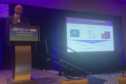
AUSTIN, TX -- Hands-on training that works better with medical students' schedules can improve practical skills and foster mentorship for promoting ultrasound use, according to research presented April 7 at UltraCon.
In his talk, Ernest Fonocho, MD, from the University of Texas Health McGovern Medical School in Houston presented findings indicating that a three-day curriculum with smaller class sizes improved image acquisition skills for students in several areas of the body.
“We think organizing these workshops is really good for medical students,” Fonocho said. “It sparks their interest in radiology and helps them learn how to use ultrasound probes, which I think most people including myself fumbled the very first time when using a probe.”
Ernest Fonocho, MD, from the University of Texas Health McGovern Medical School in Houston presents his institute's ultrasound training curriculum at UltraCon.Amerigo Allegretto
Ultrasound offers a safe, cost-effective method for real-time imaging capability. A 2021 report that Fonocho cited outlined how over 72% of medical schools indicated that they have an ultrasound curriculum. However, the report pointed out that there is a lack of formal, consistent ultrasound training.
Fonocho outlined McGovern’s previous ultrasound training method, which consisted of a week-long radiology course for second-year students. While this method provided hands-on learning and dedicated radiology and ultrasound lectures, Fonocho said it had a student-instructor ratio of 15-to-1 as well as its share of limitations. These included time constraints, a “hectic” curriculum, and inconsistent personnel availability.
The institute’s current curriculum consists of a three-day workshop during the musculoskeletal and dermatology block of the medical students’ coursework. It includes the following features: prerecorded videos introducing ultrasound, promoting independent learning prior to the workshop; radiology trainees as instructors; three workstations with ultrasound equipment and standardized patients; 50-minute learning sessions; and a student-instructor ratio ranging from four-to-one to five-to-one.
Fonocho and colleagues compared Qualtrics scores before and after the workshop for several areas of the body. These included the calcaneum, peroneal tendons, nerves, muscles, and tendons. The team found that the students’ scores improved after the three-day workshop.
| Comparison before, after 3-day ultrasound workshop | ||
|---|---|---|
| Preworkshop | Postworkshop | |
| Peroneal tendons | 42% | 47% |
| Nerves | 53% | 78% |
| Muscles | 84% | 91% |
| Tendons | 74% | 81% |
| Calcaneum | 24% | 19% |
Fonocho highlighted that the current curriculum has several benefits. These include introducing and familiarizing students to ultrasound, creating long-term interest in radiology, having radiology trainees serve as mentors, and increasing confidence in using ultrasound in clinical practice.
“Having us there and giving the students opportunities to ask questions and register for the course helps them explore radiology as an option,” Fonocho said. “We run the radiology courses for medical students, so when they meet us, it’s easier for them to approach us.”
Moving forward, Fonocho said the institute will incorporate ultrasound and imaging lectures in each standardized block during years one and two, allow time for associated interventional procedures practice by using gel models, and incorporating a longitudinal study to assess student knowledge and retention in the third and fourth years of medical student education.

![Examples of ultrasound findings and techniques. (A) Images in a 39-year-old male patient with a mass in the left thigh. The mass is heterogeneous on the B-mode US image (compared with the patient in D) and showed increased microvascularity (superb microvascular imaging [SMI]) and shear-wave elastography (SWE) values. Undifferentiated pleomorphic sarcoma was diagnosed at biopsy (with pleomorphic rhabdomyosarcoma in surgical specimen). (B) Images in an 18-year-old male patient with a mass in the left leg. The mass is hypoechoic on the B-mode image, with no other findings suggestive of malignancy. The lesion is in contact with the cortex of the tibia, which is slightly irregular. CT revealed a doubtful anteromedial tibial erosion. The microvascular study demonstrated high vascularization, suggestive of malignancy. Periosteal Ewing sarcoma was diagnosed with both histologic and immunohistochemical confirmation. (C) Images in a 69-year-old female patient with a lump growing on the outside of the left leg. Multiple SWE examinations were performed (please note the high values obtained in the measurements, whereas the color map highlights the stiffness relative to adjacent tissues). SMI showed areas of increased vascularization to target for sampling. Undifferentiated spindle cell sarcoma was diagnosed at biopsy, with residual leiomyosarcoma in the surgical specimen after neoadjuvant therapy. (D) Images in a 56-year-old female patient with a mass in the right thigh. The mass is heterogeneous at both B-mode ultrasound (similar to patient A) and MRI (coronal T2-weighted spectral attenuated inversion recovery [SPAIR]; T1-weighted pre-contrast and postcontrast imaging), which even shows uptake after the administration of paramagnetic contrast material, which is traditionally suggestive of malignancy. Low values at SMI and elastography are suggestive of benignity. Spindle cell lipoma was diagnosed at biopsy, with atypical spindle cell lipomatous tumor in the surgical specimen.](https://img.auntminnie.com/mindful/smg/workspaces/default/uploads/2026/08/images-radiol250278fig2.APCFLSvX6p.jpg?auto=format%2Ccompress&fit=crop&h=100&q=70&w=100)


![Examples of ultrasound findings and techniques. (A) Images in a 39-year-old male patient with a mass in the left thigh. The mass is heterogeneous on the B-mode US image (compared with the patient in D) and showed increased microvascularity (superb microvascular imaging [SMI]) and shear-wave elastography (SWE) values. Undifferentiated pleomorphic sarcoma was diagnosed at biopsy (with pleomorphic rhabdomyosarcoma in surgical specimen). (B) Images in an 18-year-old male patient with a mass in the left leg. The mass is hypoechoic on the B-mode image, with no other findings suggestive of malignancy. The lesion is in contact with the cortex of the tibia, which is slightly irregular. CT revealed a doubtful anteromedial tibial erosion. The microvascular study demonstrated high vascularization, suggestive of malignancy. Periosteal Ewing sarcoma was diagnosed with both histologic and immunohistochemical confirmation. (C) Images in a 69-year-old female patient with a lump growing on the outside of the left leg. Multiple SWE examinations were performed (please note the high values obtained in the measurements, whereas the color map highlights the stiffness relative to adjacent tissues). SMI showed areas of increased vascularization to target for sampling. Undifferentiated spindle cell sarcoma was diagnosed at biopsy, with residual leiomyosarcoma in the surgical specimen after neoadjuvant therapy. (D) Images in a 56-year-old female patient with a mass in the right thigh. The mass is heterogeneous at both B-mode ultrasound (similar to patient A) and MRI (coronal T2-weighted spectral attenuated inversion recovery [SPAIR]; T1-weighted pre-contrast and postcontrast imaging), which even shows uptake after the administration of paramagnetic contrast material, which is traditionally suggestive of malignancy. Low values at SMI and elastography are suggestive of benignity. Spindle cell lipoma was diagnosed at biopsy, with atypical spindle cell lipomatous tumor in the surgical specimen.](https://img.auntminnie.com/mindful/smg/workspaces/default/uploads/2026/08/images-radiol250278fig2.APCFLSvX6p.jpg?auto=format%2Ccompress&fit=crop&h=112&q=70&w=112)