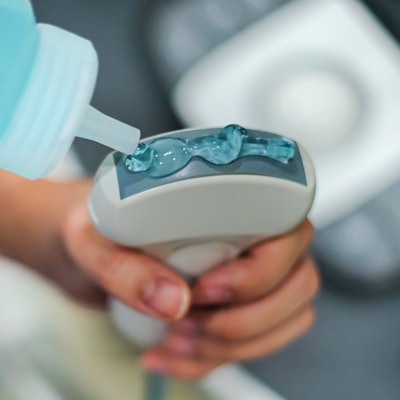
The U.S. Food and Drug Administration (FDA) said that Eco-Med Pharmaceuticals has issued a voluntary recall of certain lots of its Eco-Gel 200 ultrasound gel due to contamination from Burkholderia cepacia complex bacteria.
The bacteria causes bloodstream infections that may result in sepsis and, in certain cases, death. Use of ultrasound gels manufactured by Eco-Med have been associated with at least 15 infections to date, including bloodstream infections, the FDA statement says.
The product is also distributed as MediChoice Ultrasound Gel by Owens and Minor and Mac Medical Supply. These ultrasound gels are nonsterile and not indicated for sterile procedures, the company said.
Eco-Med is instructing all healthcare facilities to identify the affected products by lot number and immediately destroy or return products from affected lots to Eco-Med. The firm told the FDA that it's conducting a comprehensive investigation to determine the root cause of this contamination and take all necessary corrective action.
The full list of lot numbers can be found on the FDA's website.










![Examples of ultrasound findings and techniques. (A) Images in a 39-year-old male patient with a mass in the left thigh. The mass is heterogeneous on the B-mode US image (compared with the patient in D) and showed increased microvascularity (superb microvascular imaging [SMI]) and shear-wave elastography (SWE) values. Undifferentiated pleomorphic sarcoma was diagnosed at biopsy (with pleomorphic rhabdomyosarcoma in surgical specimen). (B) Images in an 18-year-old male patient with a mass in the left leg. The mass is hypoechoic on the B-mode image, with no other findings suggestive of malignancy. The lesion is in contact with the cortex of the tibia, which is slightly irregular. CT revealed a doubtful anteromedial tibial erosion. The microvascular study demonstrated high vascularization, suggestive of malignancy. Periosteal Ewing sarcoma was diagnosed with both histologic and immunohistochemical confirmation. (C) Images in a 69-year-old female patient with a lump growing on the outside of the left leg. Multiple SWE examinations were performed (please note the high values obtained in the measurements, whereas the color map highlights the stiffness relative to adjacent tissues). SMI showed areas of increased vascularization to target for sampling. Undifferentiated spindle cell sarcoma was diagnosed at biopsy, with residual leiomyosarcoma in the surgical specimen after neoadjuvant therapy. (D) Images in a 56-year-old female patient with a mass in the right thigh. The mass is heterogeneous at both B-mode ultrasound (similar to patient A) and MRI (coronal T2-weighted spectral attenuated inversion recovery [SPAIR]; T1-weighted pre-contrast and postcontrast imaging), which even shows uptake after the administration of paramagnetic contrast material, which is traditionally suggestive of malignancy. Low values at SMI and elastography are suggestive of benignity. Spindle cell lipoma was diagnosed at biopsy, with atypical spindle cell lipomatous tumor in the surgical specimen.](https://img.auntminnie.com/mindful/smg/workspaces/default/uploads/2026/08/images-radiol250278fig2.APCFLSvX6p.jpg?auto=format%2Ccompress&fit=crop&h=112&q=70&w=112)
