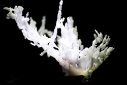
Researchers from across the U.S. confirmed the efficacy of 3D color-flow ultrasound for cheaply and reliably measuring blood flow. The findings, which were published on June 30 in Radiology, could help physicians precisely measure blood flow for people with chronic conditions.
The new approach resulted in as little as a 3%-5% difference in measurements between laboratories and operators. Based on the findings, lead study author Oliver Kripfgans, PhD, said the question of the technology's clinical adoption isn't an "if" but a "when."
"These are fantastic results that show that, from a technology point of view, some systems could be ready to go to the clinic," stated Kripfgans, an associate professor of radiology at Michigan Medicine in Ann Arbor, MI, in a press release.
2D ultrasound technology is rarely used to gather precise, quantitative blood flow measurements because results can greatly vary between facilities and operators. In addition, other noninvasive techniques to measure blood flow, such as blood pressure monitoring, can only provide qualitative data.
Kripfgans and his colleagues at Michigan Medicine had previously developed a 3D color-flow ultrasound approach to overcome the limitations of alternative measurement solutions. The researchers tested the method in the new, prospective study, which received funding from the U.S. National Institutes of Health, RSNA, and the American Institute of Ultrasound in Medicine.
For the study, Canon Medical Systems, Philips Healthcare, and GE Healthcare donated 3D ultrasound systems. The vendors modified the systems with data access software and provided the researchers with color-flow velocity, color-flow power, and scan geometry.
Trained medical or engineering staff at three different locations performed the same experimental steps using the three systems:
- They measured volume flow from 1 - 12 mL/sec in steps of 1 mL/sec at a depth of 4 cm.
- They measured depth-dependence from 2.5 - 7.5 cm in steps of 0.5 cm.
- They stepped color flow gain from no color to full blooming.
- They measured flow volume distal to a lumen stenosis. They also measured three flows at each poststenosis position.
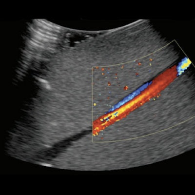
Example screenshot for poststenotic flow. Image courtesy of the RSNA.The experiments were first performed with a steady, constant flow, then repeated again with a flow simulating a pulse of 60 beats per minute. In the end, the researchers had 730 datasets with 18,450 images.

The use of 3D color-flow ultrasound produced measurements that were accurate and reproducible. Two out of the three ultrasound systems tracked within 10% for measuring flow response, the authors noted.
The approach is promising because it requires no hardware changes and can lead to practical clinical uses for measuring peripheral vascular flow and cerebral blood flow. While it will need to be further evaluated and may not work for all types of blood flow measurements, Kripfgans is optimistic about its clinical potential.
"Once the technique becomes available commercially on scanners, clinical adoption will be much faster because then it's not a research project anymore -- it's something that's readily available, and after that, it's just a matter of time before it reaches the clinic," Kripfgans stated.

![Examples of ultrasound findings and techniques. (A) Images in a 39-year-old male patient with a mass in the left thigh. The mass is heterogeneous on the B-mode US image (compared with the patient in D) and showed increased microvascularity (superb microvascular imaging [SMI]) and shear-wave elastography (SWE) values. Undifferentiated pleomorphic sarcoma was diagnosed at biopsy (with pleomorphic rhabdomyosarcoma in surgical specimen). (B) Images in an 18-year-old male patient with a mass in the left leg. The mass is hypoechoic on the B-mode image, with no other findings suggestive of malignancy. The lesion is in contact with the cortex of the tibia, which is slightly irregular. CT revealed a doubtful anteromedial tibial erosion. The microvascular study demonstrated high vascularization, suggestive of malignancy. Periosteal Ewing sarcoma was diagnosed with both histologic and immunohistochemical confirmation. (C) Images in a 69-year-old female patient with a lump growing on the outside of the left leg. Multiple SWE examinations were performed (please note the high values obtained in the measurements, whereas the color map highlights the stiffness relative to adjacent tissues). SMI showed areas of increased vascularization to target for sampling. Undifferentiated spindle cell sarcoma was diagnosed at biopsy, with residual leiomyosarcoma in the surgical specimen after neoadjuvant therapy. (D) Images in a 56-year-old female patient with a mass in the right thigh. The mass is heterogeneous at both B-mode ultrasound (similar to patient A) and MRI (coronal T2-weighted spectral attenuated inversion recovery [SPAIR]; T1-weighted pre-contrast and postcontrast imaging), which even shows uptake after the administration of paramagnetic contrast material, which is traditionally suggestive of malignancy. Low values at SMI and elastography are suggestive of benignity. Spindle cell lipoma was diagnosed at biopsy, with atypical spindle cell lipomatous tumor in the surgical specimen.](https://img.auntminnie.com/mindful/smg/workspaces/default/uploads/2026/08/images-radiol250278fig2.APCFLSvX6p.jpg?auto=format%2Ccompress&fit=crop&h=100&q=70&w=100)


![Examples of ultrasound findings and techniques. (A) Images in a 39-year-old male patient with a mass in the left thigh. The mass is heterogeneous on the B-mode US image (compared with the patient in D) and showed increased microvascularity (superb microvascular imaging [SMI]) and shear-wave elastography (SWE) values. Undifferentiated pleomorphic sarcoma was diagnosed at biopsy (with pleomorphic rhabdomyosarcoma in surgical specimen). (B) Images in an 18-year-old male patient with a mass in the left leg. The mass is hypoechoic on the B-mode image, with no other findings suggestive of malignancy. The lesion is in contact with the cortex of the tibia, which is slightly irregular. CT revealed a doubtful anteromedial tibial erosion. The microvascular study demonstrated high vascularization, suggestive of malignancy. Periosteal Ewing sarcoma was diagnosed with both histologic and immunohistochemical confirmation. (C) Images in a 69-year-old female patient with a lump growing on the outside of the left leg. Multiple SWE examinations were performed (please note the high values obtained in the measurements, whereas the color map highlights the stiffness relative to adjacent tissues). SMI showed areas of increased vascularization to target for sampling. Undifferentiated spindle cell sarcoma was diagnosed at biopsy, with residual leiomyosarcoma in the surgical specimen after neoadjuvant therapy. (D) Images in a 56-year-old female patient with a mass in the right thigh. The mass is heterogeneous at both B-mode ultrasound (similar to patient A) and MRI (coronal T2-weighted spectral attenuated inversion recovery [SPAIR]; T1-weighted pre-contrast and postcontrast imaging), which even shows uptake after the administration of paramagnetic contrast material, which is traditionally suggestive of malignancy. Low values at SMI and elastography are suggestive of benignity. Spindle cell lipoma was diagnosed at biopsy, with atypical spindle cell lipomatous tumor in the surgical specimen.](https://img.auntminnie.com/mindful/smg/workspaces/default/uploads/2026/08/images-radiol250278fig2.APCFLSvX6p.jpg?auto=format%2Ccompress&fit=crop&h=112&q=70&w=112)