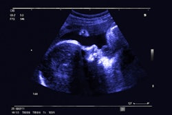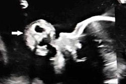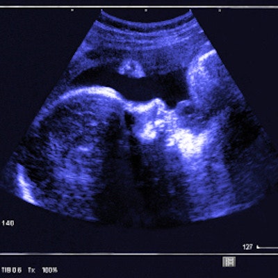
Almost a quarter of fetal abnormalities are identified for the first time during routine ultrasound at 35 to 37 weeks gestation, according to a study published on October 8 in Ultrasound in Obstetrics & Gynecology.
The study results suggest that ultrasound at this juncture of pregnancy could offer women and their babies needed support, wrote a team led by Dr. Alessandra Ficara of King's College Hospital in London.
"A high proportion of fetal abnormalities are detected for the first time during a routine ultrasound examination at 35 to 37 weeks gestation," the group wrote. "Such diagnosis and subsequent management, including selection of timing and place for delivery and postnatal investigations, could potentially improve postnatal outcome."
Assessing pregnancy at 35 to 37 weeks gestation with ultrasound helps predict the development of preeclampsia and plan delivery of a baby that is small or large for their gestational age, Ficara's team noted. But ultrasound can also identify abnormalities overlooked on early ultrasound exams.
To investigate this, Ficara and colleagues included data from 52,400 single-fetus pregnancies in women undergoing routine ultrasound exams between 35 and 37 weeks gestation.
All the women had also undergone ultrasound scans at 18 to 24 weeks of pregnancy; 47,214 had scans at 11 to 13 weeks as well. The team included pregnancies that resulted in live births or stillbirths but excluded those with known chromosomal abnormalities.
Overall, incidence of fetal abnormalities was 1.9%. Of these, 67.7% were diagnosed on ultrasound during the first or second trimester, but 24.8% were diagnosed for the first time at 35 to 37 weeks.
The most common abnormalities first identified on first- or second-trimester ultrasound but confirmed on third-trimester ultrasound included the following:
- Abdominal cyst or gastroschisis
- Cleft lip and palate
- Congenital pulmonary airway malformation
- Duplex kidney
- Hydronephrosis
- Polydactyl
- Talipes
- Unilateral multicystic kidney
- Unilateral renal agenesis and/or pelvic kidney
- Ventricular septal defect
- Ventriculomegaly
But a number of abnormalities were first identified on third-trimester ultrasound:
- Arachnoid cyst
- Duplex kidney
- Hydronephrosis
- Mild ventriculomegaly
- Ovarian cyst
- Ventricular septal defect
The study findings suggest that there's definitely a place for third-trimester ultrasound when it comes to improving pregnancy outcomes, according to the team.
"This study has highlighted the additional benefit of the late third-trimester scan in the detection of fetal abnormalities that were either missed at previous first- and second-trimester scans or became apparent only during the third trimester," the group concluded.