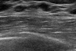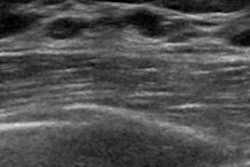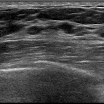
Contrast-enhanced ultrasound (CEUS) can help predict benign pathologic results of breast masses -- thus reducing the number of unnecessary biopsies by one-third, according to a study published in the September issue of the Journal of Ultrasound in Medicine.
The results could help women avoid unnecessary procedures, wrote a team led by Dr. Sandy Lee of the University of Southern California in Los Angeles.
"Our results show that CEUS can potentially be used as an additional tool in conjunction with mammography and conventional ultrasound to evaluate indeterminate BI-RADS category 4 breast masses," the group wrote. "Potentially, these masses could be downgraded to category 3 with six-month follow-up instead of biopsy."
Mammography and conventional ultrasound remain the standard imaging modalities for breast lesions, but prior studies have shown that contrast-enhanced ultrasound may help distinguish between benign and malignant breast masses, the group wrote. And although ultrasound-guided biopsies are relatively easy to perform, biopsies are invasive and expensive and can cause anxiety among patients. So Lee's team sought to investigate the use of CEUS to reduce the number of benign breast masses recommended for biopsy.
The study included 131 women with BI-RADS category 4A, 4B, and 4C masses identified on mammography, conventional ultrasound, or both between October 2016 and June 2017. The women underwent CEUS before biopsy; the researchers then compared CEUS results with histopathologic results (J Ultrasound Med, September 2019, Vol. 38:9, pp. 2259-2273).
Of the 131 masses, 109 were benign, six were high-risk, and 16 were malignant on conventional ultrasound. On CEUS, 93 of the 131 masses enhanced. Of the 109 benign lesions, 73 enhanced. All six of the high-risk lesions and 14 of the 16 malignant lesions enhanced on CEUS, the team found.
| CEUS performance in identifying breast masses | |
| Biopsy histopathology result | CEUS |
| Benign (109 masses) | 67% |
| High-risk (6 masses) | 100% |
| Malignant (16 masses) | 88% |
| Total (131 masses) | 71% |
Of the 93 masses that enhanced on CEUS, the mass shape and margin were statistically significant parameters for distinguishing between benign and malignant lesions, the researchers noted.
"Compared to high-risk and malignant masses, benign masses were more likely to have circumscribed margins when nonenhancing, oval when enhancing, and a circumscribed margin when enhancing," they wrote.
CEUS has the potential to reduce unnecessary biopsies by almost one-third, according to Lee and colleagues.
"Our study indicates that CEUS may have the potential to reduce 31% of biopsies if a mass does not enhance and has a circumscribed margin or homogeneously enhances and is oval, as these masses show benign pathologic results on tissue sampling," they concluded.


![Examples of ultrasound findings and techniques. (A) Images in a 39-year-old male patient with a mass in the left thigh. The mass is heterogeneous on the B-mode US image (compared with the patient in D) and showed increased microvascularity (superb microvascular imaging [SMI]) and shear-wave elastography (SWE) values. Undifferentiated pleomorphic sarcoma was diagnosed at biopsy (with pleomorphic rhabdomyosarcoma in surgical specimen). (B) Images in an 18-year-old male patient with a mass in the left leg. The mass is hypoechoic on the B-mode image, with no other findings suggestive of malignancy. The lesion is in contact with the cortex of the tibia, which is slightly irregular. CT revealed a doubtful anteromedial tibial erosion. The microvascular study demonstrated high vascularization, suggestive of malignancy. Periosteal Ewing sarcoma was diagnosed with both histologic and immunohistochemical confirmation. (C) Images in a 69-year-old female patient with a lump growing on the outside of the left leg. Multiple SWE examinations were performed (please note the high values obtained in the measurements, whereas the color map highlights the stiffness relative to adjacent tissues). SMI showed areas of increased vascularization to target for sampling. Undifferentiated spindle cell sarcoma was diagnosed at biopsy, with residual leiomyosarcoma in the surgical specimen after neoadjuvant therapy. (D) Images in a 56-year-old female patient with a mass in the right thigh. The mass is heterogeneous at both B-mode ultrasound (similar to patient A) and MRI (coronal T2-weighted spectral attenuated inversion recovery [SPAIR]; T1-weighted pre-contrast and postcontrast imaging), which even shows uptake after the administration of paramagnetic contrast material, which is traditionally suggestive of malignancy. Low values at SMI and elastography are suggestive of benignity. Spindle cell lipoma was diagnosed at biopsy, with atypical spindle cell lipomatous tumor in the surgical specimen.](https://img.auntminnie.com/mindful/smg/workspaces/default/uploads/2026/08/images-radiol250278fig2.APCFLSvX6p.jpg?auto=format%2Ccompress&fit=crop&h=100&q=70&w=100)


![Examples of ultrasound findings and techniques. (A) Images in a 39-year-old male patient with a mass in the left thigh. The mass is heterogeneous on the B-mode US image (compared with the patient in D) and showed increased microvascularity (superb microvascular imaging [SMI]) and shear-wave elastography (SWE) values. Undifferentiated pleomorphic sarcoma was diagnosed at biopsy (with pleomorphic rhabdomyosarcoma in surgical specimen). (B) Images in an 18-year-old male patient with a mass in the left leg. The mass is hypoechoic on the B-mode image, with no other findings suggestive of malignancy. The lesion is in contact with the cortex of the tibia, which is slightly irregular. CT revealed a doubtful anteromedial tibial erosion. The microvascular study demonstrated high vascularization, suggestive of malignancy. Periosteal Ewing sarcoma was diagnosed with both histologic and immunohistochemical confirmation. (C) Images in a 69-year-old female patient with a lump growing on the outside of the left leg. Multiple SWE examinations were performed (please note the high values obtained in the measurements, whereas the color map highlights the stiffness relative to adjacent tissues). SMI showed areas of increased vascularization to target for sampling. Undifferentiated spindle cell sarcoma was diagnosed at biopsy, with residual leiomyosarcoma in the surgical specimen after neoadjuvant therapy. (D) Images in a 56-year-old female patient with a mass in the right thigh. The mass is heterogeneous at both B-mode ultrasound (similar to patient A) and MRI (coronal T2-weighted spectral attenuated inversion recovery [SPAIR]; T1-weighted pre-contrast and postcontrast imaging), which even shows uptake after the administration of paramagnetic contrast material, which is traditionally suggestive of malignancy. Low values at SMI and elastography are suggestive of benignity. Spindle cell lipoma was diagnosed at biopsy, with atypical spindle cell lipomatous tumor in the surgical specimen.](https://img.auntminnie.com/mindful/smg/workspaces/default/uploads/2026/08/images-radiol250278fig2.APCFLSvX6p.jpg?auto=format%2Ccompress&fit=crop&h=112&q=70&w=112)