Showing people signs of atherosclerosis on their own carotid ultrasound scans can prompt them to adopt lifestyle changes that reduce their risk of cardiovascular disease, according to a new study of more than 3,500 individuals published December 3 in Lancet.
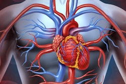
Prevention methods for cardiovascular disease often fail due to patients' low adherence to taking medication, such as statins and antihypertensive drugs, and making lifestyle changes, including those related to smoking cessation and physical activity. But new research shows the power of using personalized images of atherosclerosis as a tool to prompt behavior change and reduce the risk of cardiovascular disease, according to lead author Dr. Ulf Näslund, a professor and the chief physician in the department of public health and clinical medicine at Umeå University.
Over 3,500 patients
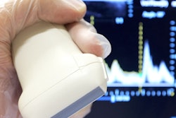
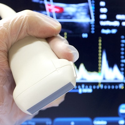
A total of 3,532 individuals who were participating in the Västerbotten County cardiovascular prevention program underwent vascular ultrasound scans of the carotid arteries. Half (1,749) were randomly selected to receive the pictorial representation of their carotid ultrasound scan, while another half (1,783) did not see their scans.
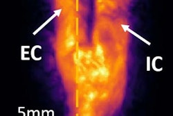
Those between 40 and 60 years old with one or more cardiovascular risk factors were eligible to participate. Participants underwent blood sampling, a survey of clinical risk factors, and an ultrasound assessment for carotid intima-media wall thickness and plaque formation.
The participants in the intervention group each received a pictorial representation of plaque formation of their arteries and also a gauge ranging from green to red to illustrate their biological age compared with their chronological age. They then received a follow-up call from a nurse after between two and four weeks to answer any questions. The same pictorial presentation of the ultrasound result was also sent to their general practitioner.
Both groups received information about their cardiovascular risk factors and a motivational health dialogue to promote healthier lifestyles, as well as pharmacological treatment according to clinical guidelines if needed.
For the one-year follow-up, the researchers calculated the cardiovascular risk scores for all participants (3,175 completed the follow-up), showing the differences between the two groups. They saw improvements for total and low-density lipoprotein cholesterol in both groups, but the team found the reduction was greater in the intervention group of patients who saw their scans versus those who didn't. The researchers also noted a graded effect, with the strongest effect seen for the individuals with most pathology on their scans.
"The differences at a population level were modest but important, and the effect was largest among those at highest risk of cardiovascular disease, which is encouraging," Näslund said in a statement. "CT and MRI might allow for a more precise assessment of risk, but these technologies have a higher cost and are not available on an equitable basis for the entire population."
"Our approach integrated an ultrasound scan, and a follow-up call with a nurse, into an already established screening program, meaning our findings are highly relevant to clinical practice," he added.
The effect of the intervention did not differ according to patients' education level, suggesting that this type of risk communication might contribute to a reduction of the social gap in health, the study authors noted. The findings come from a middle-aged population with low to moderate cardiovascular disease risk.
Looking to the future
Further research is needed to understand whether the results are sustainable beyond one year and whether showing individuals their ultrasound scans will lead to a reduction of cardiovascular disease in the long term, the authors continued. Formal cost-effectiveness analyses will be done after the three-year follow-up.
"Cardiovascular disease is the leading cause of death in many countries, and despite a wealth of evidence about effective prevention methods from medication to lifestyle changes, adherence is low," Näslund said. "Information alone rarely leads to behavior change, and the recall of advice regarding exercise and diet is poorer than advice about medicines. Risk scores are widely used, but they might be too abstract, and, therefore, fail to stimulate appropriate behaviors."
Study disclosures
Funding for the study came from the Västerbotten County Council, Swedish Research Council, Heart and Lung Foundation, Swedish Society of Medicine, and Carl Bennet, Sweden.