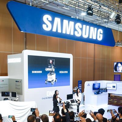
Samsung Medison, an affiliate of Samsung Electronics, is unveiling a premium ultrasound system and a prototype of a chair-type ultrasound scanner at this week's International Society of Ultrasound in Obstetrics & Gynecology (ISUOG) congress in Singapore.
A new premium platform, HERA (hyperaperture enhanced reconstruction architecture) W10 features the company's Crystal Architecture, which includes the firm's CrystalBeam beamforming technology and CrystalLive postprocessing engine. It also incorporates S-Vue transducer technology, which enables an 11-fold increase in processing power and 10 times the data transfer speed of Samsung Medison's previous system, the company said.
 The new HERA W10 ob/gyn ultrasound scanner. All images courtesy of Samsung Medison.
Other HERA W10 features include ShadowHDR, which provides a shadow-suppressed image. In addition, the company has included its HQ-Vision high-resolution imaging technology, MV-Flow blood-flow viewing technique, and LumiFlow software for 3D visualization of continuous and volumetric vascular images.
A rotatable control panel with a swivel mechanism allows for a moving range of 550 mm from left to right and 219 mm from top to bottom, Samsung Medison said.
The other new system, HERA I10, is an ergonomic chair-type ultrasound scanner that automatically adjusts for proper positioning, eliminating the need for the patient to shift the body, according to the vendor. It also reduces the force required to move the control panel back and forth and the resistance force on the wrist, the company said.
 The new HERA I10 ob/gyn ultrasound scanner on display at the ISUOG congress.
Samsung Medison is also introducing SonoSync, a software application that enables healthcare professionals to see real-time ultrasound images that are being acquired remotely. SonoSync provides two-way communication through voice chatting and marking. It can be installed on various devices, such as smartphones, laptops, and desktop PCs, facilitating real-time training and education among healthcare professionals at multiple locations, according to the firm.