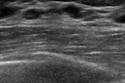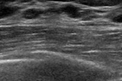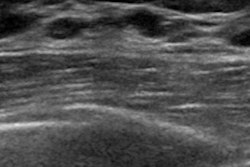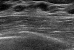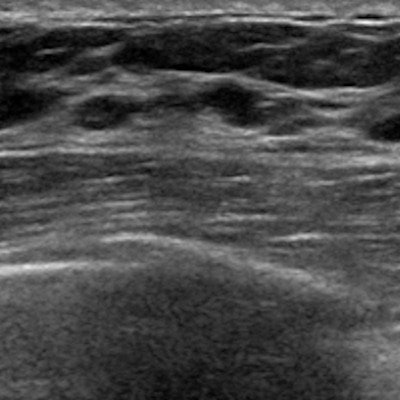
Should the current BI-RADS 3 ultrasound category -- covering breast lesions that are "probably benign" -- be revised? It appears so, according to research published online October 17 in Ultrasound in Medicine and Biology.
Although studies have shown that lesions in the ultrasound BI-RADS 3 category pose a malignancy risk of less than 2% -- and therefore can be safely followed in the short term with further imaging -- not everyone agrees that this is the best way to track them, wrote a team led by Dr. Chiara Pistolese from the University of Rome Tor Vergata. In fact, some practices recommend that women with BI-RADS 3 lesions undergo core-needle or surgical biopsy, just to be sure.
"Breast ultrasound, in daily practice, is an indispensable and irreplaceable method in breast nodule detection and characterization," the group wrote. "The introduction of the BI-RADS system helped radiologists to describe the sonographic characteristics and to assign lesions to a category associated with the most appropriate clinical treatment. Despite the help offered by this classification system, the BI-RADS 3 category includes probably benign lesions whose management has been widely debated in the literature."
Pistolese and colleagues sought to evaluate the validity of the BI-RADS 3 category in ultrasound by using cytologic and histologic results as the reference standard. The researchers included 122 patients with 122 nodular lesions found on breast ultrasound and classified as BI-RADS 3, all of which were biopsied percutaneously (Ultrasound Med Biol, October 17, 2018).
In 86 cases (70.5%), the biopsies showed benign results; 29 cases (23.7%) had uncertain results and seven (5.7%) had malignant results. Some of these malignant lesions had cysts or small anechoic components that have typically been associated with benign lesions, according to Pistolese's team.
The presence of these typically benign characteristics in malignant lesions -- as well as the higher than expected malignancy rate -- surprised the researchers and led them to believe that the BI-RADS 3 category for ultrasound may need to be revised.
"The management of probably benign nodular lesions should not only be guided by BI-RADS classification; it is also necessary to include clinical ... data and apply a multidisciplinary approach to select cases that require histologic verification instead of the usual follow-up," the group wrote.
"Despite the small cohort, our data seem to indicate the need to revise the current BI-RADS 3 category," the authors concluded. "It could be useful to evaluate as suspicious the presence of nonhomogeneous echoes within the corpuscular cyst and solid nodular lesions with cystic components."


![Examples of ultrasound findings and techniques. (A) Images in a 39-year-old male patient with a mass in the left thigh. The mass is heterogeneous on the B-mode US image (compared with the patient in D) and showed increased microvascularity (superb microvascular imaging [SMI]) and shear-wave elastography (SWE) values. Undifferentiated pleomorphic sarcoma was diagnosed at biopsy (with pleomorphic rhabdomyosarcoma in surgical specimen). (B) Images in an 18-year-old male patient with a mass in the left leg. The mass is hypoechoic on the B-mode image, with no other findings suggestive of malignancy. The lesion is in contact with the cortex of the tibia, which is slightly irregular. CT revealed a doubtful anteromedial tibial erosion. The microvascular study demonstrated high vascularization, suggestive of malignancy. Periosteal Ewing sarcoma was diagnosed with both histologic and immunohistochemical confirmation. (C) Images in a 69-year-old female patient with a lump growing on the outside of the left leg. Multiple SWE examinations were performed (please note the high values obtained in the measurements, whereas the color map highlights the stiffness relative to adjacent tissues). SMI showed areas of increased vascularization to target for sampling. Undifferentiated spindle cell sarcoma was diagnosed at biopsy, with residual leiomyosarcoma in the surgical specimen after neoadjuvant therapy. (D) Images in a 56-year-old female patient with a mass in the right thigh. The mass is heterogeneous at both B-mode ultrasound (similar to patient A) and MRI (coronal T2-weighted spectral attenuated inversion recovery [SPAIR]; T1-weighted pre-contrast and postcontrast imaging), which even shows uptake after the administration of paramagnetic contrast material, which is traditionally suggestive of malignancy. Low values at SMI and elastography are suggestive of benignity. Spindle cell lipoma was diagnosed at biopsy, with atypical spindle cell lipomatous tumor in the surgical specimen.](https://img.auntminnie.com/mindful/smg/workspaces/default/uploads/2026/08/images-radiol250278fig2.APCFLSvX6p.jpg?auto=format%2Ccompress&fit=crop&h=100&q=70&w=100)


![Examples of ultrasound findings and techniques. (A) Images in a 39-year-old male patient with a mass in the left thigh. The mass is heterogeneous on the B-mode US image (compared with the patient in D) and showed increased microvascularity (superb microvascular imaging [SMI]) and shear-wave elastography (SWE) values. Undifferentiated pleomorphic sarcoma was diagnosed at biopsy (with pleomorphic rhabdomyosarcoma in surgical specimen). (B) Images in an 18-year-old male patient with a mass in the left leg. The mass is hypoechoic on the B-mode image, with no other findings suggestive of malignancy. The lesion is in contact with the cortex of the tibia, which is slightly irregular. CT revealed a doubtful anteromedial tibial erosion. The microvascular study demonstrated high vascularization, suggestive of malignancy. Periosteal Ewing sarcoma was diagnosed with both histologic and immunohistochemical confirmation. (C) Images in a 69-year-old female patient with a lump growing on the outside of the left leg. Multiple SWE examinations were performed (please note the high values obtained in the measurements, whereas the color map highlights the stiffness relative to adjacent tissues). SMI showed areas of increased vascularization to target for sampling. Undifferentiated spindle cell sarcoma was diagnosed at biopsy, with residual leiomyosarcoma in the surgical specimen after neoadjuvant therapy. (D) Images in a 56-year-old female patient with a mass in the right thigh. The mass is heterogeneous at both B-mode ultrasound (similar to patient A) and MRI (coronal T2-weighted spectral attenuated inversion recovery [SPAIR]; T1-weighted pre-contrast and postcontrast imaging), which even shows uptake after the administration of paramagnetic contrast material, which is traditionally suggestive of malignancy. Low values at SMI and elastography are suggestive of benignity. Spindle cell lipoma was diagnosed at biopsy, with atypical spindle cell lipomatous tumor in the surgical specimen.](https://img.auntminnie.com/mindful/smg/workspaces/default/uploads/2026/08/images-radiol250278fig2.APCFLSvX6p.jpg?auto=format%2Ccompress&fit=crop&h=112&q=70&w=112)