South Korean researchers found that by replacing an interventional needle with a catheter, they were better able to perform interventional radiology treatment of endometrial cysts under ultrasound guidance. They described the technique in a study published August 28 in Radiology.
The procedure not only offers a less-invasive alternative to surgery -- which can negatively affect fertility -- it is also more effective than needle-directed sclerotherapy, wrote a team led by Dr. Kichang Han of Yonsei University College of Medicine in Seoul.
"Surgical excision has been considered the standard treatment for ovarian endometrioma," the group wrote. "However, a decline in the ovarian reserve after surgical cystectomy is inevitable due to removal of healthy ovarian tissue adjacent to the endometrioma and/or excessive electrocoagulation to the ovary for hemostasis. This outcome has prompted a shift toward less-invasive approaches."
Improving the alternative
Endometriosis is a condition in which the lining of the uterus grows into surrounding tissues and organs. It affects about 10% of women in their reproductive years, and when it involves the ovaries, cysts called endometriomas can form, often leading to pelvic pain and uterine bleeding.
Ultrasound-guided sclerotherapy has been used as an alternative to surgery since 1988, primarily with needles, Han and colleagues wrote. But this approach has limitations, including a lack of stability of the needle during aspiration of the cyst and a risk of needle displacement. Han's team developed catheter-directed sclerotherapy to address these drawbacks.
"Endometrial cyst content can be incredibly thick and sticky," said study co-author Dr. Man-Deuk Kim, PhD, in a statement released by the RSNA. "A 16- or 18-gauge needle, which is commonly used for needle-directed sclerotherapy, is sometimes not large enough to completely evacuate the cyst. This may reduce the efficacy of sclerotherapy and increase the possibility of needle displacement during aspiration."
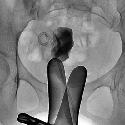
Catheter-directed sclerotherapy involves an interventional radiologist inserting a catheter rather than a needle under fluoroscopy guidance into the endometrioma to drain it. The radiologist then fills the cyst with contrast to determine whether any of its content has spilled into the pelvic cavity. The contrast is aspirated and ethanol injected into the cyst. After 20 minutes, the ethanol is removed and the catheter taken out.
The study included 14 women who had the procedure between March 2015 and December 2017. The researchers assessed the technique's impact on the women's ovaries by measuring serum anti-Müllerian hormone (AMH), an indicator of ovarian health, before and after the procedure and six months later. The group also measured serum cancer antigen 125 (CA-125) levels at these same time points to assess the procedure's effectiveness. Finally, study participants underwent follow-up ultrasound at one, three, and six months after catheter-directed sclerotherapy and biannually thereafter to track cyst size changes and/or recurrence.
Overall, the women tolerated the procedure well, their intraprocedural pain was managed effectively, and there were no complications. At the six-month follow-up, there were no cyst recurrences and no statistically significant differences in AMH levels, and CA-125 levels decreased. Among the 14 participants, two desired pregnancy; both succeeded in becoming pregnant after the procedure.
| Patient outcomes with catheter-directed sclerotherapy | ||
| Before therapy | 6 months after therapy | |
| Mean cyst size | 5.8 cm | 1.1 cm |
| Pain score | 3.86 | 0.6 |
| Ovarian reserve (AMH) | 4.29 ng/mL | 4.36 ng/mL* |
| CA-125 | 65.4 U/mL | 20.9 U/mL |
 Clinical course of a 27-year-old woman with primary ovarian endometrioma presenting with dysmenorrhea. (a) Axial T2-weighted image reveals an 8.4-cm, multilocular endometrioma (*) with internal septation (arrows). (b) The endometrioma was punctured and mechanical breakdown of the internal septa was performed. (c) A catheter was inserted and contrast was injected to observe any leakage. (d) Follow-up ultrasound six months after catheter-directed sclerotherapy shows markedly decreased endometrioma (*). Images and captions courtesy of RSNA.
"In our series, there were no short-term recurrences, and a significant decrease in serum CA-125 level was observed," the group wrote. "In addition, 100% of participants experienced alleviation of various pain symptoms."
Effective treatment
Catheter-directed sclerotherapy is an effective variation of the needle-directed approach, Han and colleagues wrote.
"With several technical advantages, catheter-directed sclerotherapy seems to be a promising modification of needle-directed sclerotherapy to manage ovarian endometrioma," they concluded. "Moreover, ovarian reserve was well-preserved in patients who underwent catheter-directed sclerotherapy for endometriomas, which may contribute to future fertility."