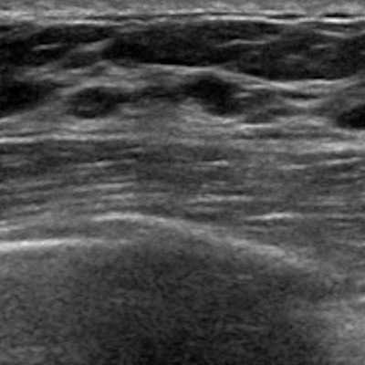
Breast density notification laws have increased the use of follow-up ultrasound after mammography screening, according to a study published online July 20 in Medical Care.
Women with denser breast tissue are considered to have a higher risk for breast cancer, in part due to a masking effect this type of tissue can have on lesions and also the decreased sensitivity of mammography in this population, wrote a team led by Michal Horný, PhD, of Emory University in Atlanta. In response to a grassroots effort on the part of patient advocates, more than 30 states have passed laws mandating that women be notified if they have dense tissue and advised of adjunct imaging options.
"By 2018, 31 states had enacted legislation in response to issues in mammography screening in women with increased breast density," Horný said in a statement released by the Harvey L. Neiman Health Policy Institute, which supported the research. "Of the 31 states ... 20 did so before the end of 2014. We identified characteristics of policies implemented in these 20 states from the published literature and a review of the enacted bills."
Horný's group used a sample of 13.4 million screening mammography exams acquired from the MarketScan Research database. The exams were performed between 2007 and 2014 on privately insured women in states that had implemented breast density notification legislation during that period. The researchers found that notification of patients about issues surrounding increased breast density was associated with increased ultrasound follow-up by 1.02 percentage points.
The effect of these laws is likely particular to the approaches taken by individual states, the researchers concluded.
"The heterogeneous effect of state legislation with regard to dense breast tissue on screening mammography follow-up by ultrasound may be explained by specific and unique characteristics of the approaches taken by a variety of states," they wrote. "Some policy characteristics, such as the explicit suggestion of supplemental imaging or mandated coverage of supplemental imaging by health insurance, augmented that effect."










![Examples of ultrasound findings and techniques. (A) Images in a 39-year-old male patient with a mass in the left thigh. The mass is heterogeneous on the B-mode US image (compared with the patient in D) and showed increased microvascularity (superb microvascular imaging [SMI]) and shear-wave elastography (SWE) values. Undifferentiated pleomorphic sarcoma was diagnosed at biopsy (with pleomorphic rhabdomyosarcoma in surgical specimen). (B) Images in an 18-year-old male patient with a mass in the left leg. The mass is hypoechoic on the B-mode image, with no other findings suggestive of malignancy. The lesion is in contact with the cortex of the tibia, which is slightly irregular. CT revealed a doubtful anteromedial tibial erosion. The microvascular study demonstrated high vascularization, suggestive of malignancy. Periosteal Ewing sarcoma was diagnosed with both histologic and immunohistochemical confirmation. (C) Images in a 69-year-old female patient with a lump growing on the outside of the left leg. Multiple SWE examinations were performed (please note the high values obtained in the measurements, whereas the color map highlights the stiffness relative to adjacent tissues). SMI showed areas of increased vascularization to target for sampling. Undifferentiated spindle cell sarcoma was diagnosed at biopsy, with residual leiomyosarcoma in the surgical specimen after neoadjuvant therapy. (D) Images in a 56-year-old female patient with a mass in the right thigh. The mass is heterogeneous at both B-mode ultrasound (similar to patient A) and MRI (coronal T2-weighted spectral attenuated inversion recovery [SPAIR]; T1-weighted pre-contrast and postcontrast imaging), which even shows uptake after the administration of paramagnetic contrast material, which is traditionally suggestive of malignancy. Low values at SMI and elastography are suggestive of benignity. Spindle cell lipoma was diagnosed at biopsy, with atypical spindle cell lipomatous tumor in the surgical specimen.](https://img.auntminnie.com/mindful/smg/workspaces/default/uploads/2026/08/images-radiol250278fig2.APCFLSvX6p.jpg?auto=format%2Ccompress&fit=crop&h=112&q=70&w=112)


