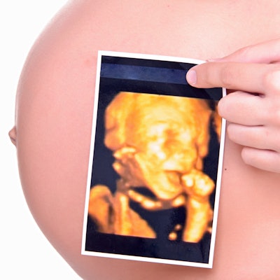
A study published in February in JAMA Pediatrics that suggested a link between the incidence of autism spectrum disorder (ASD) and prenatal ultrasound is coming under fire by critics who are calling out the study's conclusion as oversimplified at best and inaccurate at worst.
The original research, conducted by a team led by Dr. N. Paul Rosman of Boston Medical Center, claimed to find a statistically significant association between the development of ASD and one technical sonography parameter: the depth of ultrasound penetration for scans performed at certain points during pregnancy.
But this measure isn't valid, wrote Dr. Christoph Lees from the Imperial College Healthcare NHS Trust in London in a letter published online June 11, also in JAMA Pediatrics.
"The authors introduce a hitherto unheard-of index: the depth of ultrasonographic penetration," he wrote. "This has no clear biological or scientific justification. Nevertheless, they report that the mean depth of penetration of the ultrasound beam is greater in those who developed ASD than in healthy children. But depth of penetration of ultrasound is not a measure of ultrasonography exposure; it simply tells us how far the ultrasound beam reaches."
The study authors' conclusion that "further research is needed to determine whether other variables of ultrasound exposure also have adverse effects on the developing fetus" doesn't accurately reflect the data, he wrote.
"In apparent contravention of JAMA Pediatrics' guidance for reporting clinical studies of this nature, no recruitment flowchart with participant exclusion/inclusion criteria is shown, and neither a priori hypothesis, predefined primary outcome, nor sample size calculation are included," Lees wrote.
The study also drew criticism in a letter from Drs. David Somerset and Robert Wilson of the University of Calgary, who believe the study's conclusion that "greater ultrasonographic depth" negatively affects the fetus has not been proved and is a "gross oversimplification."
"The authors present no evidence that a mean difference of 4 mm in depth is associated with higher energy delivery to the fetus," they wrote. "Furthermore, clinical variables, such as maternal obesity and age and paternal age, are independently associated with ASD ... and these variables have not been accounted for. It is a shame that ... [the] study has such an alarmist conclusion in the abstract that is not supported by the study findings."
Because possible links between ultrasound and autism cause such anxiety among parents and pregnant women, it's crucial that studies are reported accurately, Lees concluded.
"In fact, this study shows that children with ASD were exposed to a shorter duration of prenatal ultrasonography and lower-energy scans than healthy children," he wrote. "These findings are reassuring. We respectfully suggest that a more accurate form of words would have reflected the study's finding than is found in the conclusion of the abstract."