Handheld-ultrasound technology developer Clarius Mobile Health is touting the use of its scanner for a novel application: patrolling trails at a mountain resort in Canada to diagnose skiing and mountain biking injuries.
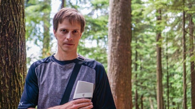
Dr. Geoff Sanz is an emergency physician who also works as a patrol hut doctor at a resort in Kelowna, British Columbia. He carries the company's pocket-sized Clarius scanner with him on his travels around the resort, using it to evaluate injured skiers, snowboarders, and mountain bikers.
 Dr. Geoff Sanz hits the trail with the Clarius handheld scanner. Photo courtesy of Clarius.
The resort is over a mile from the nearest hospital, so being able to render a rapid diagnosis in the field is critical. The scanner helps Sanz diagnose not only those whose injuries require ambulance or air transfer, but also those with low-risk injuries who can make it to the hospital in their own vehicle.
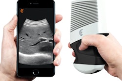
The Clarius scanner operates on the iOS and Android platforms and is powered by a rechargeable battery that can be exchanged when running low. The system also comes in a magnesium case that enables it to withstand challenging environments. It is also water-submersible for easy cleaning and disinfection.







![Examples of ultrasound findings and techniques. (A) Images in a 39-year-old male patient with a mass in the left thigh. The mass is heterogeneous on the B-mode US image (compared with the patient in D) and showed increased microvascularity (superb microvascular imaging [SMI]) and shear-wave elastography (SWE) values. Undifferentiated pleomorphic sarcoma was diagnosed at biopsy (with pleomorphic rhabdomyosarcoma in surgical specimen). (B) Images in an 18-year-old male patient with a mass in the left leg. The mass is hypoechoic on the B-mode image, with no other findings suggestive of malignancy. The lesion is in contact with the cortex of the tibia, which is slightly irregular. CT revealed a doubtful anteromedial tibial erosion. The microvascular study demonstrated high vascularization, suggestive of malignancy. Periosteal Ewing sarcoma was diagnosed with both histologic and immunohistochemical confirmation. (C) Images in a 69-year-old female patient with a lump growing on the outside of the left leg. Multiple SWE examinations were performed (please note the high values obtained in the measurements, whereas the color map highlights the stiffness relative to adjacent tissues). SMI showed areas of increased vascularization to target for sampling. Undifferentiated spindle cell sarcoma was diagnosed at biopsy, with residual leiomyosarcoma in the surgical specimen after neoadjuvant therapy. (D) Images in a 56-year-old female patient with a mass in the right thigh. The mass is heterogeneous at both B-mode ultrasound (similar to patient A) and MRI (coronal T2-weighted spectral attenuated inversion recovery [SPAIR]; T1-weighted pre-contrast and postcontrast imaging), which even shows uptake after the administration of paramagnetic contrast material, which is traditionally suggestive of malignancy. Low values at SMI and elastography are suggestive of benignity. Spindle cell lipoma was diagnosed at biopsy, with atypical spindle cell lipomatous tumor in the surgical specimen.](https://img.auntminnie.com/mindful/smg/workspaces/default/uploads/2026/08/images-radiol250278fig2.APCFLSvX6p.jpg?auto=format%2Ccompress&dpr=2&fit=crop&h=167&q=70&w=250)


