The long wait is over. The U.S. Food and Drug Administration (FDA) for the first time has approved the use of an ultrasound contrast agent for radiology applications, giving the nod to Lumason from Bracco Diagnostics to be used for focal liver lesions.
Lumason was known previously in the U.S. as SonoVue, and it received initial FDA approval in October 2014 for use in patients with suboptimal echocardiograms to opacify the left ventricular chamber and to improve the delineation of the left ventricular endocardial border. The agent is still offered by Bracco under the SonoVue brand outside of the U.S.
Notably, the FDA's approval this week of a supplemental new drug application (sNDA) for Lumason for liver applications covers both adult and pediatric patients, said Dr. Alberto Spinazzi, senior vice president of global medical and regulatory affairs at Bracco Group. One phase II and two phase III clinical trials were used to support the indication for characterizing focal liver lesions in adult patients, while published data in the literature were used to support the use of Lumason in pediatric patients, he said.
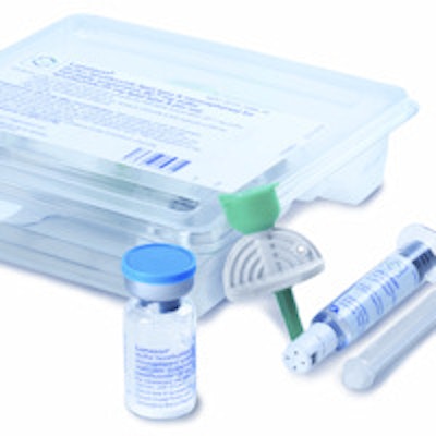
 Lumason ultrasound contrast from Bracco.
Liver imaging has been an established indication for ultrasound contrast in several countries in Europe and in Asia. The approval of Lumason in the U.S. now for this application is clinically important, as is the approved use in children, Spinazzi said.
With the long wait for FDA approval for radiology indications, many in the ultrasound contrast community have begun over the past few years to publicly advocate the off-label use of these agents. However, it's still important that the FDA has approved Lumason for this liver indication, Spinazzi said.
"We believe it is important that something that was used off-label now is in some way validated by the FDA," he told AuntMinnie.com. "The review of the FDA is extensive; they review all of the evidence and they assess what is the risk-benefit for a new use of a product. We believe FDA review is critical to ensuring the safety and efficacy of any use of imaging products to the healthcare community."
Safety concerns involving the use of ultrasound contrast agents had led the FDA to issue a black box safety warning in 2007. While lobbying efforts led to the FDA later relaxing that black box warning, it still remains in place for all three ultrasound contrast agents currently approved in the U.S.: Lumason from Bracco, Optison from GE Healthcare, and Definity from Lantheus Medical Imaging.
Even when the FDA was focused on the safety issues involved with ultrasound contrast, the agency has always been open to approving new indications for ultrasound contrast, Spinazzi said.
"They have considered not only the evidence that was related specifically to the use of the product in liver imaging, but all of the safety information available on the agent -- cardiac, noncardiac, everything," he said. "In the end, they provided this approval."