Diffractive ultrasound (DUS) is a sonographic technique that shows promise for breast imaging, according to researchers at the 2003 congress of the World Federation for Ultrasound in Medicine and Biology and the American Institute of Ultrasound in Medicine in Montreal.
"It is especially good at identifying solid lesions," said Dr. Beverly Hashimoto, a radiologist from Virginia Mason Medical Center in Seattle. "And ductal and lobular carcinomas show up particularly well."
DUS combines two ultrasound beams to form a 3-D holographic pattern of the breast. In a study (funded by Advanced Imaging Technologies of Preston, WA, which recently filed for Chapter 11 bankruptcy protection and is being reorganized), Hashimoto and her colleague Dr. Lynn Wiitala found DUS and mammography to be superior to conventional sonography and mammography for identifying solid breast masses, but inferior for detecting cystic lesions.
"Solid-lesion detection is more critical when it comes to identifying cancer," said Wiitala, who was the principal investigator.
The researchers imaged 129 lesions in 125 patients. DUS with mammography identified 39 out of 55 (70.9%) cystic lesions, compared to 100% detection by conventional ultrasound and mammography.
However, for solid lesions, DUS and mammography detected 47 out of 51 (92.2%). In comparison, conventional ultrasound and mammography detected 40 out of 51 (80.4%).
For malignant solid masses, DUS and mammography had the same detection rate as conventional ultrasound and mammography at 91.7%. Mammography alone had a lower detection rate at 75%.
Hashimoto proposed a set of DUS imaging terms that she hoped would foster better communication of findings between investigators using different imaging methods, and would also help incorporate DUS into the complement of breast imaging techniques.
Categories of the proposed classification system include descriptions of a lesion’s shape, margin, and absorption, as well as indicators of normal and abnormal breast architecture, she said.
DUS uses a gray scale that correlates white with high sound transmission and dark with low sound transmission, making cysts look relatively white and solid masses look dark.
 |
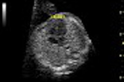
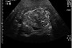
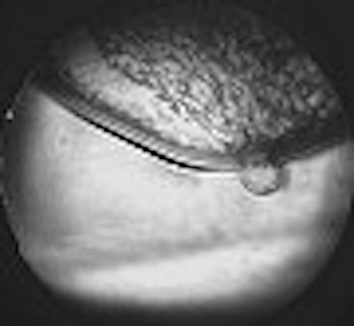
A fibroademona (above) and a ductal carcinoma (middle) as seen on diffractive ultrasound. Below, normal findings that highlight the different tissue layers in the breast. Images courtesy of Advanced Imaging Technologies.
  |
On DUS, cystic lesions are round, oval, or lobular in shape, with circumscribed margins. Solid masses are oval, lobulated, or irregular in shape, with either circumscribed or ill-defined margins. Malignant tumors exhibit architectural distortion by distorting the pattern produced by the surrounding fibroglandular tissue.
"It’s a promising technique insofar as differentiating cysts and solid masses. If it’s dark on DUS we know it’s solid. But whether it’s benign or not we can’t tell," Hashimoto said.
By Kate JohnsonAuntMinnie.com contributing writer
July 18, 2003
Related Reading
Sydney team reiterates value of US for younger breasts, June 10, 2003
US plus mammo characterizes palpable breast lesions, May 30, 2003
Breast ultrasound firm AIT files for bankruptcy, April 25, 2003
Strict attention to detail enhances sensitivity of breast US, April 17, 2003
Copyright © 2003 AuntMinnie.com