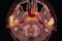
Researchers from Vanderbilt University have developed PET imaging agents that could make it possible to visualize tumors in their earliest stages before they become deadly, according to a study in the October issue of Cancer Prevention Research.
The compounds, derived from inhibitors of the enzyme cyclooxygenase-2 (COX-2) and detectable by PET, may have broad applications for cancer detection, diagnosis, and treatment.
Lead study author Lawrence Marnett, PhD, director of the Vanderbilt Institute of Chemical Biology, said the compound is the first COX-2-targeted PET imaging agent validated for use in animal models of inflammation and cancer (Cancer Prev Res, October 2011, Vol. 4:10, pp. 1536-1545).
COX-2 is not found in most normal tissues but is activated in inflammatory lesions and tumors. As a tumor grows and becomes increasingly malignant, COX-2 levels increase.
Researchers incorporated radioactive fluorine into the compound, and intravenous injection of the compound into animal models provided a sufficient signal to be detected by PET.
The results support further development of the agents as probes for early detection of cancer and for evaluating the COX-2 status of premalignant and malignant tumors, Marnett and colleagues concluded.
The research was supported by the U.S. National Cancer Institute (NCI).


