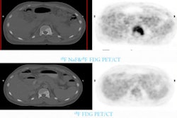
For the first time, quantitative, rather than qualitative, data analysis has demonstrated that time-of-flight (TOF) PET scans can improve cancer detection, according to a new study in the March issue of the Journal of Nuclear Medicine.
The research from Harvard Medical School and Massachusetts General Hospital (MGH) found that oncologic TOF FDG-PET scans produced significant improvements in lesion detection of lung and liver cancers over all contrasts and body mass indexes.
The lead author of the study is Georges El Fakhri, PhD, from the division of nuclear medicine and molecular imaging at Harvard and MGH.
Conventional PET scans create images by detecting gamma rays produced by radioisotopes that are injected into the body. However, the scans do not consider the time for each gamma ray to reach the detector. TOF PET scans do calculate travel time, which results in enhanced image signal-to-noise.
In the study, TOF PET images were compared to conventional PET images to determine if there were any improvements in lesion detection relative to location, scan time, contrast, and body mass index.
Improved lesion detection was observed in the TOF PET scans, with the greatest gains in the shortest-acquisition studies and in the subjects with a body mass index of 30 or greater. In addition, the greatest gain in performance was achieved at the lowest lesion contrast and the smallest gain in performance at the highest lesion contrast.
The researchers concluded that nuclear medicine technologists and physicians may be able to take advantage of the gain achieved with TOF PET to reduce scanning time, increase patient comfort, and minimize patient motion.
They may also be able to reduce the injected radiopharmaceutical dose, decreasing radiation exposure to patients and healthcare professionals.