Nuclear medicine has seen explosive growth in cardiac imaging since the 1990s, with much of the expansion driven by the migration of nuclear cardiology studies from hospital settings and into the offices of cardiologists who have purchased their own gamma cameras. The cardiology office segment is continuing to show strength, and it's attracting a number of start-up firms that have developed dedicated cardiac cameras optimized for in-office imaging.
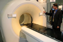
One of those firms is Israeli gamma camera developer Spectrum Dynamics. The Tirat Hacarmel firm exhibited at its first Society of Nuclear Medicine (SNM) show this month with its D-SPECT gamma camera, a dedicated cardiac system that the company intends to launch in early 2007. The system was first demonstrated at the 2005 RSNA show.
Spectrum Dynamics developed its gamma camera design with the goal of addressing what the company believes are two major issues in nuclear cardiology imaging in physician offices: patient throughput and limitations on existing gamma camera design leading to the inability to do absolute quantification of blood-flow data.
For the first issue, as patient studies have moved to cardiologist offices, many practices have developed huge patient backlogs, according to Josh Gurewitz, marketing consultant for the firm. "We realized that one of the key drivers in this space is throughput," Gurewitz said. "We needed to be able to do things faster without sacrificing image quality, and if possible improve image quality, which would improve diagnostic accuracy."
Spectrum Dynamics decided to jettison the conventional Anger-based design that has been at the core of gamma cameras for the past five decades. In its place, the company developed a technology it calls BroadView, which houses 10 cadmium zinc telluride solid-state digital detectors in an L-shaped arc around the patient, who sits in a chair during the study.
Rather than rotating the detector array around the patient, or the patient around the detector array, the system swivels each detector back and forth during the study, with the range of motion automatically based on the patient's body mass index and a scout view scan. This calculation enables D-SPECT to spend more time imaging the heart, with fewer photon counts coming from structures outside the organ, Gurewitz said.
 |
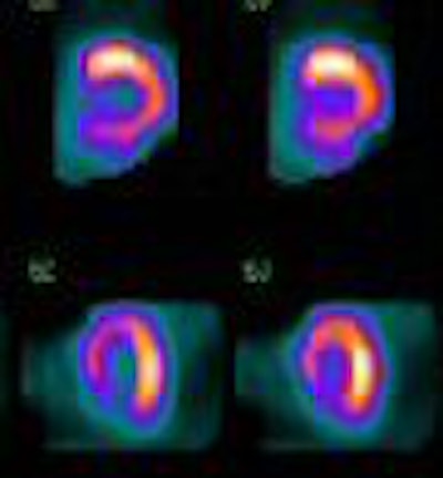
| Tc-99m sestamibi rest myocardial perfusion study of 57-year-old woman with body mass index of 33. Risk factors included hypertension, smoking, and family history of coronary artery disease. Echocardiography revealed mild left ventricular enlargement, normal ejection fraction, hypokinesis of the distal lateral wall, mitral valve prolapse, and mitral insufficiency. Image collected in two-minute gated acquisition with D-SPECT system. |
Spectrum Dynamics calls the concept of setting imaging parameters automatically based on patient physiology and anatomy "personalized molecular scanning." It's a similar idea to the approach being taken in the pharmaceutical industry.
"One protocol doesn't fit all," Gurewitz said. "Patients come in different sizes and shapes, so we optimize (scanning parameters) based on their anatomy and physiology."
D-SPECT can conduct a standard gated SPECT study in two minutes, and in the future the company plans to enable the system to conduct a stress/rest simultaneous gated SPECT study with technetium sestamibi and thallium in eight minutes -- major improvements on cardiac cameras with standard Anger detector technology, Gurewitz said.
But Spectrum Dynamics didn't just stop with D-SPECT's instrumentation. The company developed an entire workflow process based on reducing medical errors and speeding patients through the imaging exam. When patients are checked in, the receptionist downloads all relevant patient and procedure data from the facility's information system to a radiofrequency ID (RFID) chip. The RFID chip is attached to a wristband that is then given to the patient to wear.
When it's time for the injection of the radiopharmaceutical, the information on the RFID chip on the wristband that indicates what procedure the patient is scheduled to receive must match up with the RFID chip on the plunger of the radiopharmaceutical syringe. If it doesn't, the technologist will receive a warning stating that the two are not a match and that the dose should not be administered, Gurewitz said.
When the patient sits down at the D-SPECT camera, the system uploads patient information to the camera's acquisition module wirelessly, helping avoid the redundant input of patient information and improving workflow. The RFID data also helps the system track the correct radiopharmaceutical dose and calculate the scanning parameters needed to personalize the study.
Spectrum Dynamics demonstrated the system at the SNM show as a work-in-progress, and hopes to have D-SPECT in beta testing by September, with commercial shipments starting in the first quarter of 2007 pending U.S. Food and Drug Administration clearance.
Four U.S. sites that are luminary nuclear cardiology centers will be among the initial beta sites, including Cedars-Sinai Medical Center in Los Angeles, Brigham and Women's Hospital in Boston, Vanderbilt Medical Center in Nashville, and Baptist Cardiac and Vascular Institute of Miami. The firm expects that the system will carry a list price competitive with existing fixed-detector 90° cardiac cameras.
By 2008, Spectrum Dynamics hopes to be able to offer a version of D-SPECT in a SPECT/CT configuration. The company is working on a design that would enable the SPECT and CT modules to be detached from each other and operated independently, which would improve workflow and provide clinical flexibility for facilities that have installed the technology, Gurewitz said.
Another possible future application is absolute quantification, in which back-to-back fast SPECT acquisitions are conducted, with the system producing temporal resolution that's high enough to offer absolute quantification of blood flow down to a resolution of several seconds. The company believes this would be an improvement over current cardiac imaging methods that use relative quantification, Gurewitz said.
By Brian Casey
AuntMinnie.com staff writer
June 19, 2006
Related Reading
Spectrum Dynamics shows D-SPECT system, June 7, 2006
Copyright © 2006 AuntMinnie.com