Dear AuntMinnie Member,
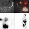
The Society of Nuclear Medicine this week gave its Image of the Year honor to a cardiac image created through a fusion of SPECT and CT angiography studies.
Continuing an SNM tradition, Dr. Henry Wagner made the announcement on Monday at the society's annual meeting in San Diego. Staff writer Jonathan S. Batchelor was on hand to report for our Molecular Imaging Digital Community
The winning image was produced by researchers at University Hospital Zurich in Switzerland using a 64-slice CT scanner and a dual-head SPECT system, with image fusion performed using postprocessing software. The study demonstrates how anatomical heart imaging modalities like CTA can benefit from the functional information added by SPECT and PET, Dr. Wagner said. See the image for yourself by clicking here.
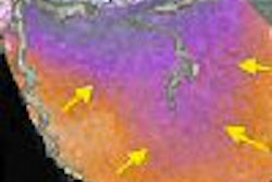
We're also featuring other news from SNM, including a study on the use of whole-body PET/CT for colorectal cancer staging, available here, and a presentation on dual-tracer PET for imaging cerebral gliomas, available here. Another study discussed the development of a computer-aided detection algorithm to assess interval changes on bone scintigraphy studies, which you can read about by clicking here.
In related news, we're highlighting excerpts from Dr. Wagner's newly released book, A Personal History of Nuclear Medicine: Reflections of a Pioneer, in which he looks back on the milestones of his illustrious 50-year career in nuclear medicine. Just click here for fascinating highlights from Dr. Wagner's new book.
You'll find all these stories, plus additional coverage from the SNM show, in our Molecular Imaging Digital Community: molecular.auntminnie.com.