It may be the greatest thing since sliced bread in other applications. But when faced with the diagnostic challenge presented by the undisplaced scaphoid fracture, 16-slice multidetector CT apparently doesn't make the cut.
Not that the cut is clearly defined. There is no universally accepted gold standard for the diagnosis, and thus no way to truly gauge the modality's accuracy in this area.
But based on a head-to-head comparison with skeletal scintigraphy, British researchers have reluctantly concluded that MDCT probably shouldn't be the next step after initial x-rays fail to show a suspected wrist fracture.
The latest study was led by Ashley M. Groves from the radiology department at Addenbrooke's Hospital and the University of Cambridge in England and published in the American Journal of Roentgenology (AJR, May 2005, Vol. 184:5, pp. 1470-1474).
As described by the authors, commonly occurring scaphoid fractures have long posed a clinical challenge. Physical exams are unreliable and radiographs aren't generally regarded as definitive in ruling out an undisplaced fracture.
MRI stands out among the tested modalities, which include SPECT and ultrasound. "Some institutions use MRI as a primary investigation in the emergency department for suspected radiologically occult wrist fracture," the authors noted.
But the cost of and access to MRI may be an assumed barrier in some regions. Meanwhile, scaphoid fractures should be identified early, the authors noted, "as immediate treatment is required to minimize the chances of nonunion and avascular necrosis."
Inspired by the imaging advances incorporated in MDCT, including improved visualization of bone cortex and trabecular patterns, the researchers proceeded to compare MDCT scans with skeletal scintigraphy in 51 patients.
All of the patients had presented to the trauma center after an injury, but had no fracture detected on x-rays taken immediately or at follow-up 10 to 14 days later. All patients subsequently underwent MDCT scanning on the same day as their skeletal scintigraphy exam.
Scintigraphy consisted of planar images of the wrists obtained three hours after the injection of 400 MBq of technetium-99m-methylene diphosphonate (99mTc-MDP).
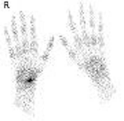
 |
| Nineteen-year-old man with wrist pain after fall. Local-view 99mTc-MDP skeletal scintigram of wrist shows focal uptake of tracer in right scaphoid. R = right. |
CT images were acquired with 0.75-mm detectors and reconstructed in 0.5-mm increments. Fracture was diagnosed when a cortical breech was visualized.
Of the 51 patients examined, 23 were diagnosed with fractures on scintigraphy while only 14 were deemed to have fractures on CT. Of the nine additional fractures identified by scintigraphy, five involved the scaphoid.
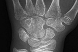
 |
| Nineteen-year-old man with wrist pain after fall. Coronal CT reconstruction of wrist shows no evidence of fracture. Image has been reversed to match image above. Groves AM, Cheow H, Balan K, Courtney H, Bearcroft P, and Dixon A, "16-MDCT in the Detection of Occult Wrist Fractures: A Comparison with Skeletal Scintigraphy" (AJR 2005; 184:1470-1474). |
Four of the discrepant cases involved patients with arthritic changes that would have made CT diagnosis more difficult, although arthropathy might also confound some scintigraphic interpretations as well, the authors noted.
"The fact that scintigraphy appeared to detect more fractures than CT is both intuitive and in keeping with the current literature," the authors wrote. "However, the use of CT has some inherent advantages. Patients appear to prefer it, no needles are involved, and examination lengths were about 7 minutes."
On the other hand, CT required more time for interpretation and management of the vast set of images produced.
"Despite some inherent advantages of 16-MDCT, our findings make it difficult to recommend CT as an initial investigation in suspected scaphoid fracture," the authors concluded. "CT will probably be useful, however, in cases of equivocal scintigraphic findings or when needed to visualize the fracture line, such as to assess healing."
"The finding that some patients have positive scintigraphy results but normal results on CT and six-weeks' follow-up radiography is interesting and challenges our understanding of patients presumed to have a fracture on the basis of skeletal scintigraphy," the authors continued. "Further research -- for example, with MRI -- is warranted."
By Tracie L. Thompson
AuntMinnie.com staff writer
May 18, 2005
Related Reading
MRI and CT: Complementary techniques in assessing rheumatoid arthritis, March 9, 2005
Panoramic x-ray improves detection of challenging wrist fractures, January 8, 2004
The twists and turns of hand and wrist x-ray positioning, October 15, 2002
Ultrasound detects occult scaphoid fractures, June 26, 2002
Earlier is better for MRI in wrist trauma, February 10, 2002
Copyright © 2005 AuntMinnie.com