
Prerecorded, available throughout meeting | SPR-MS-13
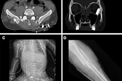
Using an abbreviated whole-body trauma CT protocol is an effective way to streamline imaging in a mass-casualty event when emergency rooms may be swamped, researchers have found.The findings could translate to better patient care in a stressful situation, wrote a team led by presenter Dr. Muhammad Israr Ahmad of the University of British Columbia in Vancouver.
"In a mass casualty incident, CT is an important tool for triage but can be a bottleneck to patient care," Ahmad's team wrote in the study abstract. "Improved scan efficiency allows more rapid patient throughput, helping more patients be assessed/treated."
Ahmad's group developed a shortened "disaster" whole-body CT protocol as an alternative to conventional whole-body CT. The abbreviated protocol included axial CT head, axial CT angiogram vertex to pelvis, and sagittal reformat cervical spine. Four emergency radiologists read only these images from 10 whole-body CT complex trauma exams.
The readers identified 92% of acute traumatic findings; of these, 97% were identified by staff radiologists and 87% by fellows. There were some missed findings, but they were low grade and would not necessarily require immediate treatment in a mass-casualty event.
Experience did affect the performance of readers, Ahmad and colleagues discovered: Fellows were more likely to miss findings, while false positives were more likely among staff radiologists.
Yes, an abbreviated whole-body CT protocol may miss some low-grade conditions, but in a mass-casualty event, it appears to be an effective way to streamline imaging, the team concluded.
"Use of an abbreviated whole body trauma CT protocol in a mass casualty incident can expedite imaging without missing those injuries that require immediate treatment," Ahmed and colleagues wrote.


![Axial images from unenhanced calcium score cardiac CT (left) and curved planar reformation images from CT angiography (right) show that higher long-term exposure to air pollution is associated with greater coronary artery calcium and more obstructive coronary artery disease (CAD). Top row: Images in a 68-year-old male patient with higher 10-year mean ambient air pollution exposure (7.9 μg/m3 for particulate matter measuring ≤2.5 μm in diameter [PM2.5] and 17.4 parts per billion [ppb] for NO2) with extensive CAD (coronary artery calcium score [CACS] >1,000 and obstructive CAD [≥70% diameter stenosis]). Bottom row: Images in a 57-year-old female patient with lower 10-year mean ambient air pollution exposure (6.3 μg/m3 for PM2.5 and 4.6 ppb for NO2) with no CAD (CACS = 0 and no obstructive stenosis).](https://img.auntminnie.com/mindful/smg/workspaces/default/uploads/2026/06/hanneman.r6SMLzkezo.png?auto=format%2Ccompress&dpr=2&fit=crop&h=167&q=70&w=250)


