Research using CT to image a 25-million-year-old fossil whale skull has shown that Aetiocetus weltoni -- a distant relation to today's Mysticeti baleen whales -- had teeth and baleen simultaneously in adulthood.
The combination made for a crowded mouth, according to the researchers from San Diego State University (SDSU) and the San Diego Natural History Museum. The study findings illuminate the transition from teeth to baleen in these ancient creatures and were published May 24 in the Zoological Journal of the Linnean Society.
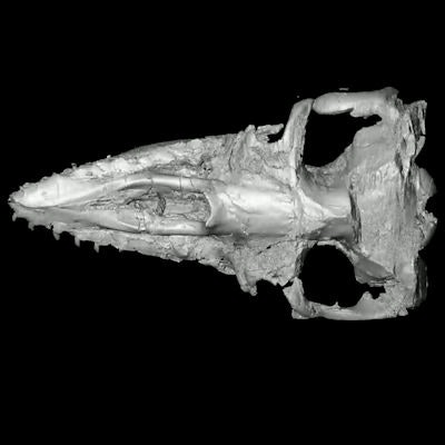
 3D digital reconstruction of Aetiocetus weltoni skull. Image courtesy of Eric Ekdale, SDSU.
The team led by Eric Ekdale, PhD, of SDSU used CT imaging to investigate evidence of baleen in Aetiocetus. The study showed "grooves and holes on the roof of the mouth that connect internally with a vascular canal in a fashion consistent with the pattern of blood vessels that lead to baleen in modern mysticetes," SDSU noted in a statement.
The findings reveal that the blood supply for the teeth was diverted to support the growth of baleen.
"Our study provides tangible fossil evidence of a major shift in feeding behavior from a raptorial carnivorous feeding mode to a bulk filter-feeding mode for obtaining food, among the largest animals that have ever lived in Earth's oceans," Ekdale said.