CT can effectively define the scope of trauma in pediatric bicycle handlebar injuries and help clinicians decide on the best treatment, according to a study published May 4 in Radiographics.
If the full impact of bicycle handlebar injuries isn't understood, it may not be clear what kind of treatment is appropriate, wrote a team led by Dr. Deepa Reddy Biyyam of Phoenix Children's Hospital. That's where CT can help -- and why radiologists are so important for assessing these injuries.
"The radiologist plays an important role in the workup and treatment of patients with handlebar injuries," the team wrote. "CT remains the preferred examination to help diagnose chest and abdominal trauma, with higher sensitivity and specificity when compared to those of ultrasound and clinical findings. Understanding of imaging findings and patterns ... helps clinicians in deciding on surgical or conservative management."
Nonmotorcycle bicycle handlebar injuries are a prominent cause of chest and abdominal trauma in children, but they have often been overlooked in clinical research, according to Biyyam and colleagues. The injuries result in about "430,000 hospital visits and 275 deaths annually in the U.S. ," and in one study, "50% of children who sustained handlebar trauma experienced life-threatening intra-abdominal injury," the group wrote. But physical findings may not be definitive.
"The physical findings are often underwhelming, and laboratory values in many studies are shown to be not very sensitive or specific," the authors wrote. "As a result, there is a risk of delay in imaging, diagnosis, and treatment of significant and sometimes life-threatening injuries. CT is considered the standard examination to delineate intra-abdominal trauma ... [and] helps in grading some types of injury and ... [guiding] the surgical treatment course."
Biyyam's team listed the following injuries caused by handlebar trauma that can be diagnosed with CT and described some of the findings' characteristics.
- Thoracic: Rib fractures, pneumothorax, pulmonary contusion, pneumomediastinum
- Abdominal: Pancreas, small bowel, mesentery, liver, spleen, adrenal gland (liver lacerations may present as linear hypoattenuating lesions; spleen lacerations range from linear to branching patterns)
- Pancreatic: Contusion and inflammation ("focal or diffuse enlargement of the pancreas with decreased enhancement and associated peripancreatic fat stranding and fluid")
- Duodenal: Hematomas (may visualize as "an intraluminal, intramural, or paraduodenal collection with increased attenuation"), perforations, lacerations ("interruption of the enhancing bowel wall, extravasation of intraluminal contents, or free retroperitoneal air")
- Abdominal wall hernia ("usually found at weak anatomic locations such as lower abdomen lateral to the rectus sheath")
- Groin and vascular ("CT angiography can help identify occlusion of the artery")
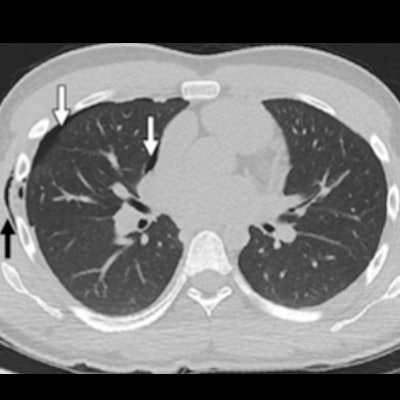
 Chest wall injury in a 16-year-old adolescent boy with a direct handlebar injury to the right chest. (a) Axial CT image of the chest (lung window) demonstrates soft-tissue emphysema in the right lateral chest wall (black arrow) with a small right pneumothorax (white arrows). (b) Axial CT image (bone window) demonstrates a nondisplaced right sixth rib fracture (arrow). Images courtesy of the RSNA.
 Splenic laceration in a 10-year-old child with a handlebar injury to the left upper quadrant. Axial (a) and coronal (b) contrast-enhanced CT images show a large disruption of the spleen (arrow) extending into the splenic hilum, a finding consistent with a grade 4 splenic laceration. Perisplenic free fluid and hemorrhage are also noted.
It's hard to overstate how important it is to work up bicycle handlebar injuries in children, according to the researchers.
"Given the often subtle injuries and the significant potential morbidity related to handlebar trauma, it is crucial for every radiologist to be aware of the patterns of handlebar injuries for timely diagnosis and management," they concluded.