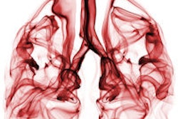
Patients with nonsolid lung nodules can be safely managed noninvasively using low-dose CT, according to an analysis published online June 23 in Radiology of more than 57,000 lung cancer screening patients.
The findings could help spare patients from unnecessary surgery and additional imaging, reported Dr. Claudia Henschke, PhD, from Weill Cornell Medical College, and colleagues.
Nonsolid nodules can represent inflammation, infection, or fibrosis in addition to cancer; therefore, in lung cancer screening there is a need to define which nodules need invasive workup and which can be managed noninvasively, as well as how often the follow-up should occur, the study team wrote.
From the original International Early Lung Cancer Action Program (I-ELCAP) cohort of 57,496 patients, Henschke and colleagues identified nonsolid nodules in 2,392 (4.2%) of the baseline screening exams. Further analysis revealed 73 cases of cancer (Radiology, June 23, 2015).
The researchers also detected new nonsolid nodules in 485 (0.7%) of 64,677 annual repeat scans, and 11 patients were diagnosed with stage I cancer. However, all surgery was curative at a median follow-up of six years after diagnosis.
Nonsolid nodules developed a solid component, indicating a heightened risk of invasive cancer, in 22 cases, but the median transition time from nonsolid to part-solid was more than two years. No cancer was found in new nodules 15 mm or larger.
The results suggest that nonsolid nodules can be followed safely with low-dose CT at 12-month intervals, regardless of size, to assess potential transitions to part-solid nodules, Henschke and colleagues concluded.
The findings can help reduce unnecessary CT scans and even biopsies and surgeries in lung cancer screening programs, according to the group.

![Images show the pectoralis muscles of a healthy male individual who never smoked (age, 66 years; height, 178 cm; body mass index [BMI, calculated as weight in kilograms divided by height in meters squared], 28.4; number of cigarette pack-years, 0; forced expiratory volume in 1 second [FEV1], 97.6% predicted; FEV1: forced vital capacity [FVC] ratio, 0.71; pectoralis muscle area [PMA], 59.4 cm2; pectoralis muscle volume [PMV], 764 cm3) and a male individual with a smoking history and chronic obstructive pulmonary disorder (COPD) (age, 66 years; height, 178 cm; BMI, 27.5; number of cigarette pack-years, 43.2, FEV1, 48% predicted; FEV1:FVC, 0.56; PMA, 35 cm2; PMV, 480.8 cm3) from the Canadian Cohort Obstructive Lung Disease (i.e., CanCOLD) study. The CT image is shown in the axial plane. The PMV is automatically extracted using the developed deep learning model and overlayed onto the lungs for visual clarity.](https://img.auntminnie.com/mindful/smg/workspaces/default/uploads/2026/03/genkin.25LqljVF0y.jpg?auto=format%2Ccompress&crop=focalpoint&fit=crop&h=100&q=70&w=100)


![Images show the pectoralis muscles of a healthy male individual who never smoked (age, 66 years; height, 178 cm; body mass index [BMI, calculated as weight in kilograms divided by height in meters squared], 28.4; number of cigarette pack-years, 0; forced expiratory volume in 1 second [FEV1], 97.6% predicted; FEV1: forced vital capacity [FVC] ratio, 0.71; pectoralis muscle area [PMA], 59.4 cm2; pectoralis muscle volume [PMV], 764 cm3) and a male individual with a smoking history and chronic obstructive pulmonary disorder (COPD) (age, 66 years; height, 178 cm; BMI, 27.5; number of cigarette pack-years, 43.2, FEV1, 48% predicted; FEV1:FVC, 0.56; PMA, 35 cm2; PMV, 480.8 cm3) from the Canadian Cohort Obstructive Lung Disease (i.e., CanCOLD) study. The CT image is shown in the axial plane. The PMV is automatically extracted using the developed deep learning model and overlayed onto the lungs for visual clarity.](https://img.auntminnie.com/mindful/smg/workspaces/default/uploads/2026/03/genkin.25LqljVF0y.jpg?auto=format%2Ccompress&crop=focalpoint&fit=crop&h=112&q=70&w=112)


