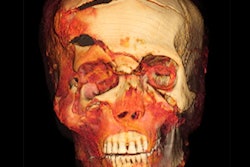
Cincinnati Children's Hospital said that it used x-ray, CT, and 3D printing to learn new information about a 500-year-old child mummy from Peru.
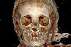
From the imaging, radiologists from the institution determined that the mummy was a 2- to 3-year-old girl at the time of her death. After also discovering a small hole pierced through the child's spine, they created digital 3D models and collaborated with the hospital's department of clinical engineering to create a 3D printed model in order to get a better understanding of the lesion.
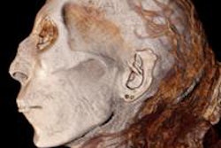
After evaluating the imaging and the 3D models as well as consulting with scientific researchers associated with the exhibit, the group determined that the hole represented a sampling of the mummy that occurred after it was collected, according to the hospital. The 3D printed model also showed the abnormal shape of the child's head, which was consistent with the common practice at the time of aesthetic molding for beautification purposes, the institution said.
While the researchers were unable to determine the cause of death, they were able to rule out several possibilities, including tuberculosis, major trauma to the body, widespread cancer, thyroid dysfunction, or some other abnormality that would cause death, the hospital said. The radiographs also revealed growth-recovery lines in the child's arms and legs, indicating a nutritional stress that had stopped growth for a period of time.
The mummy, which was brought to the hospital as part of a scientific project with the Cincinnati Museum Center, is currently on display in the museum's "Mummies of the World: The Exhibition" until April.