Sunday, November 25 | 11:15 a.m.-11:25 a.m. | SSA18-04 | Room S505AB
FDG-PET/CT may be used as primary staging modality in patients with primary breast cancer, according to this Sunday morning presentation to be given by German researchers.Dr. Till Heusner, from the University of Düsseldorf, and colleagues compared the diagnostic accuracy of whole-body FDG-PET/CT for initial breast cancer staging with the accuracy of a conventional, multimodality imaging algorithm. The team also sought to explore whether patient management would be changed due to the FDG-PET/CT findings.
The study included 106 breast cancer patients who had whole-body FDG-PET/CT and also conventional imaging (x-ray mammography, breast MRI, chest x-ray, bone scintigraphy, and breast, axillary, and liver ultrasound). Heusner's group evaluated diagnostic accuracy by primary tumor detection rate, correct assessment of primary lesion location, T stage, and the detection rates of lymph node and distant metastases.
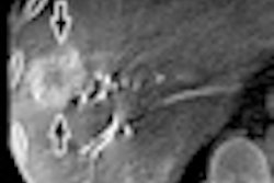
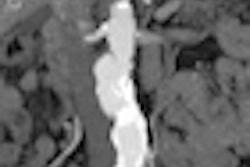
FDG-PET/CT was more accurate for detecting axillary lymph node and distant metastases, the group found: In fact, in 14 patients, only FDG-PET/CT found synchronous tumors, extra-axillary lymph node metastases, or distant metastases. Fourteen percent of patients had their disease management plans changed because of FDG-PET/CT findings -- a substantial proportion, the researchers concluded.