Computer-aided detection (CAD) firm iCAD of Nashua, NH, plans to debut a new version of its VeraLook CAD software for CT colonography (CTC) at the European Society of Gastrointestinal and Abdominal Radiology (ESGAR) annual meeting in Dresden, Germany, in June.
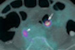
VeraLook is available in Europe under CE Mark approval; in the U.S., it is pending clearance by the U.S. Food and Drug Administration (FDA). The software detects and highlights potential polyps during CTC examinations, and the newest version features performance improvements including a greater than 5% increase in the sensitivity of potential polyp detection and a 20% reduction in false positives, according to iCAD.
Bichat Hospital in Paris has recently installed VeraLook for CTC and is using it with a V3D workstation from Viatronix of Stony Brook, NY, iCAD said.
Related Reading
iCAD revenue slips in Q1, April 29, 2010
iCAD supports educational sessions, March 16, 2010
iCAD sales dip as firm posts profit, February 23, 2010
iCAD partners with AdMeTech, November 18, 2009
iCAD to show CAD for tomo at RSNA, November 13, 2009
Copyright © 2010 AuntMinnie.com


![Images show the pectoralis muscles of a healthy male individual who never smoked (age, 66 years; height, 178 cm; body mass index [BMI, calculated as weight in kilograms divided by height in meters squared], 28.4; number of cigarette pack-years, 0; forced expiratory volume in 1 second [FEV1], 97.6% predicted; FEV1: forced vital capacity [FVC] ratio, 0.71; pectoralis muscle area [PMA], 59.4 cm2; pectoralis muscle volume [PMV], 764 cm3) and a male individual with a smoking history and chronic obstructive pulmonary disorder (COPD) (age, 66 years; height, 178 cm; BMI, 27.5; number of cigarette pack-years, 43.2, FEV1, 48% predicted; FEV1:FVC, 0.56; PMA, 35 cm2; PMV, 480.8 cm3) from the Canadian Cohort Obstructive Lung Disease (i.e., CanCOLD) study. The CT image is shown in the axial plane. The PMV is automatically extracted using the developed deep learning model and overlayed onto the lungs for visual clarity.](https://img.auntminnie.com/mindful/smg/workspaces/default/uploads/2026/03/genkin.25LqljVF0y.jpg?auto=format%2Ccompress&crop=focalpoint&fit=crop&h=100&q=70&w=100)


![Images show the pectoralis muscles of a healthy male individual who never smoked (age, 66 years; height, 178 cm; body mass index [BMI, calculated as weight in kilograms divided by height in meters squared], 28.4; number of cigarette pack-years, 0; forced expiratory volume in 1 second [FEV1], 97.6% predicted; FEV1: forced vital capacity [FVC] ratio, 0.71; pectoralis muscle area [PMA], 59.4 cm2; pectoralis muscle volume [PMV], 764 cm3) and a male individual with a smoking history and chronic obstructive pulmonary disorder (COPD) (age, 66 years; height, 178 cm; BMI, 27.5; number of cigarette pack-years, 43.2, FEV1, 48% predicted; FEV1:FVC, 0.56; PMA, 35 cm2; PMV, 480.8 cm3) from the Canadian Cohort Obstructive Lung Disease (i.e., CanCOLD) study. The CT image is shown in the axial plane. The PMV is automatically extracted using the developed deep learning model and overlayed onto the lungs for visual clarity.](https://img.auntminnie.com/mindful/smg/workspaces/default/uploads/2026/03/genkin.25LqljVF0y.jpg?auto=format%2Ccompress&crop=focalpoint&fit=crop&h=112&q=70&w=112)


