The Medical Imaging and Technology Alliance (MITA) has endorsed eight principles to reduce exposure to unnecessary medical radiation, minimize medical errors, and improve reporting of adverse events, the Arlington, VA-based organization said.
The proposals come amid heightened scrutiny of radiation from medical sources. The U.S. Food and Administration (FDA) this week announced that it plans a new initiative to more closely regulate diagnostic and therapeutic radiation, while the U.S. Congress is also expected to hold hearings on the issue.
In its proposal, MITA recommends:
- Expanding and integrating appropriateness criteria into physician decision-making
- Creating a national dosage registry to ensure longitudinal tracking of dose levels for patients across America
- Adopting a standardized method of storing diagnostic imaging and radiation therapy information within electronic health records
- Expanding mandatory accreditation for advanced imaging facilities
- Establishing minimum standards for training and education for hospital and imaging facility personnel who perform medical imaging exams and deliver radiation therapy treatments
- Developing enhanced operational safety procedures and checklists to reduce medical errors
- Expanding and standardizing the reporting of medical errors associated with medical radiation across stakeholders in a manner that is transparent for patients, families, and physicians
- Developing radiation dose reference values to provide a data point to compare the dose level of a specific procedure
Related Reading
MITA plans rally to defend medical imaging, October 14, 2009
NEMA names new MITA head, September 24, 2009
MITA names new managing director, December 4, 2008
NEMA releases MRI standards, October 28, 2008
MITA's Whitman joins Varian, July 29, 2008
Copyright © 2010 AuntMinnie.com


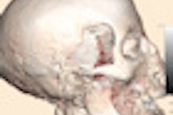
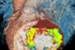


![Axial images from unenhanced calcium score cardiac CT (left) and curved planar reformation images from CT angiography (right) show that higher long-term exposure to air pollution is associated with greater coronary artery calcium and more obstructive coronary artery disease (CAD). Top row: Images in a 68-year-old male patient with higher 10-year mean ambient air pollution exposure (7.9 μg/m3 for particulate matter measuring ≤2.5 μm in diameter [PM2.5] and 17.4 parts per billion [ppb] for NO2) with extensive CAD (coronary artery calcium score [CACS] >1,000 and obstructive CAD [≥70% diameter stenosis]). Bottom row: Images in a 57-year-old female patient with lower 10-year mean ambient air pollution exposure (6.3 μg/m3 for PM2.5 and 4.6 ppb for NO2) with no CAD (CACS = 0 and no obstructive stenosis).](https://img.auntminnie.com/mindful/smg/workspaces/default/uploads/2026/06/hanneman.r6SMLzkezo.png?auto=format%2Ccompress&dpr=2&fit=crop&h=167&q=70&w=250)


