Mesenchymal tumors are a heterogeneous group of intramural lesions that arise deep to the mucosa.
1.4 Mesenchymal Tumors
CLINICAL FEATURES
• Mesenchymal tumors are a heterogeneous group of intramural lesions that arise deep to the mucosa.
• Gastrointestinal stromal tumor (GIST) accounts for the majority of solid mesenchymal neoplasms; these tumors arise from the interstitial cells of Cajal (GI pacemaker cells).
• Other mesenchymal tumors include smooth muscle tumors (e.g., leiomyoma), neurogenic tumors (e.g., schwannoma, neurofibroma), vascular tumors (e.g., hemangioma), and hamartomas.
• GISTs were previously misclassified as smooth muscle tumors.
• Mesenchymal tumors may present with acute or occult GI bleeding secondary to ulceration of the overlying mucosa.
• Malignant mesenchymal tumors typically spread via direct local extension, intraperitoneal seeding, or hematogenously (to liver).
• Small bowel lipomas are generally benign incidental findings but may lead to intussusception.
• Hamartomas consist of varying amounts of smooth muscle and epithelial tissue.
• Multiple small bowel hamartomas are a common finding in Peutz-Jeghers syndrome; these benign polyps may act as lead points for intussusception.
• Hemangiomas and lymphangiomas of the small bowel are usually asymptomatic incidental findings; see section 3.9 for discussion of vascular ectasia.
• Most nerve sheath tumors (i.e., schwannomas and neurofibromas) are benign.
IMAGING FEATURES
• Mesenchymal tumors are intramural masses that typically manifest as smooth, broad-based impressions on barium studies; nonulcerated lesions may be difficult to distinguish from extrinsic impression.
• CT and MR are useful for demonstrating both the intramural and exoenteric tumor components, which are typically much more prominent than the endoluminal component.
• CT can evaluate for metastatic disease and monitor response to therapy (which is often striking in cases of GIST).
• A densely calcified lesion is suggestive of a leiomyoma.
• CT can provide an imaging-specific diagnosis for lipomas, obviating tissue sampling.
• Hemangiomas are typically small but can present as large polypoid lesions; the presence of phleboliths may allow for a specific diagnosis.
• Lymphangiomas may present as a polypoid or lobulated submucosal lesion and may be pliable at fluoroscopy and endoscopy; most will appear cystic on cross-sectional imaging.
FIGURES
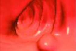
Figure 1.4.1 Small bowel GIST.
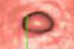
Figure 1.4.2 Ulcerated small bowel GIST.
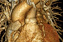
Figure 1.4.3 Small bowel lipomas.
Figure 1.4.4 Small bowel hamartomas.
Figure 1.4.5 Peutz-Jeghers syndrome.
Copyright © 2007 by Saunders, an imprint of Elsevier, Inc.