Computer-aided detection (CAD) developer R2 Technology has launched its ImageChecker CT Lung Version 2.0 CAD software system and AutoPoint temporal comparison feature for use during review of multidetector-row CT chest exams.
The latest iteration of the visualization application features enhancements to R2's CT lung CAD algorithm as well as expanded PACS integration, according to the Sunnyvale, CA-based company.
An AutoPoint temporal comparison option uses automatic 3D registration to enable physicians to track nodule changes over time, R2 said.
The system also provides a tabular report featuring key images and information on nodule volume and density changes, as well as a nodule's estimated growth doubling time, a measure of the likelihood of the nodule being malignant, the developer said.
By AuntMinnie.com staff writers
May 17, 2005
Related Reading
R2 adds to executive lineup, May 13, 2005
iCAD, R2 in patent dispute, April 28, 2005
Vital Images, R2 form alliance, April 26, 2005
R2 gets FDA nod for CAD with Mammomat, February 23, 2005
R2, FirstChoice contract extended, February 10, 2005
Copyright © 2005 AuntMinnie.com


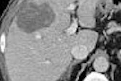
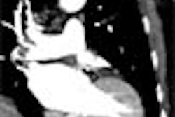


![Axial images from unenhanced calcium score cardiac CT (left) and curved planar reformation images from CT angiography (right) show that higher long-term exposure to air pollution is associated with greater coronary artery calcium and more obstructive coronary artery disease (CAD). Top row: Images in a 68-year-old male patient with higher 10-year mean ambient air pollution exposure (7.9 μg/m3 for particulate matter measuring ≤2.5 μm in diameter [PM2.5] and 17.4 parts per billion [ppb] for NO2) with extensive CAD (coronary artery calcium score [CACS] >1,000 and obstructive CAD [≥70% diameter stenosis]). Bottom row: Images in a 57-year-old female patient with lower 10-year mean ambient air pollution exposure (6.3 μg/m3 for PM2.5 and 4.6 ppb for NO2) with no CAD (CACS = 0 and no obstructive stenosis).](https://img.auntminnie.com/mindful/smg/workspaces/default/uploads/2026/06/hanneman.r6SMLzkezo.png?auto=format%2Ccompress&dpr=2&fit=crop&h=167&q=70&w=250)


