Dear AuntMinnie Member,
The buzz was certainly back at RSNA 2023. Attendance was noticeably higher at McCormick Place, as radiology’s premier event continues to bounce back from pandemic levels.
AI once again took center stage in Chicago, both in the scientific sessions and in the technical exhibits. This year, there was a noticeable emphasis on back-end applications such as image reconstruction and on helping radiologists become more efficient by streamlining reporting, for example.
Although largely not yet ready for clinical use, generative AI is also showing potential for tasks such as integrating information from prior exams and in patient engagement. Continuing a recent trend, vendors are also increasingly developing platforms aimed at easing integration of AI apps into radiology workflows.
Looking to catch up on all of the key research and industry news from the meeting? Check out our RSNA 2023 RADcast, which was updated daily throughout the week with articles and video interviews with key luminaries.
Our Top 5 articles so far from RSNA 2023 are as follows:
- DTI-MRI reveals impact of soccer heading on brain
- Change needed for radiology to grow
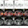
- PET tracer shows promise in prostate cancer patients
- Video from RSNA 2023: AI and Fight Club
- Elastography predicts treatment response in colorectal cancer patients
Erik L. Ridley
Editor in Chief
AuntMinnie.com